
10-step guide


Radiation Therapy
Treatment planning, biological modelling , image guided radiotherapy.
Multi-centre/international comparisons
Clinical trials outcomes
At UWA, Medical Physics research is strongly aligned with improving treatment and diagnostic precision using localized and minimally invasive techniques that are aimed to improve patient outco mes.
Studying Medical Physics at UWA provides an exceptional opportunity to learn in a department with strong commitment to research. The research component of this programme allows students to develop valuable skills for practising and interpreting research.
Research opportunities are available for students. To submit an expression of interest for a research opportunity, fill out our form
Radiation Therapy (RT)
Biological modelling of tumour and normal tissue dose responses
Modelling the interaction of radiotherapy and immunotherapy
Intensity Modulated Radiation Therapy (IMRT)
3D printing application in radiation therapy
Intra-Operative Radiation Therapy (IORT)
Image Guided Radiation Therapy (IGRT)
Modelling, Simulation, and Prototyping
Development of monitoring devices
Assessment of impact of inaccuracy
Treatment planning optimization
Stereotactic Radiosurgery (SRS)
Tracking patient/organ motion
Treatment delivery accuracy
QA on treatment equipment
Small-animal radiotherapy
Robotics applications
Pre-clinical studies
Immunotherapy
3-D dosimetry
Drug studies
Nuclear Medicine (NM)
Implementation of a SPECT quality phantom QA regime in a nuclear medicine department
Creation of semi-automated image analysis tools for PET processing
Image analysis, dosimetry and theranostics radiopharmaceuticals
Radiation Biology (RB)
Bio-guided radiotherapy planning
Simulation of cancer tissue
Simulation of treatment effect
Optimisation of treatment
Patient-specific simulation
Radiation Protection (RP) and Medical Health Physics (MHP)
Identification and Quantification of Radioisotopes with a HPGe Gamma Spectrometer
Diagnostic Imaging (DI)
Realistic breast models for optimising electromagnetic gradiometric measurements
Molecular imaging
Bio-imaging with magnetic gradiometry
Other themes
Biostatistics
3D data processing
Machine learning methods
Medical Physics Research
The mission of physics research is to develop new techniques and investigate new methodologies to continuously improve treatment planning and evaluation methods, target localization and verification accuracy and treatment delivery precision.
Please click below to learn more about physics research.
Investigators
Qiuwen Wu, Zheng (Jim) Chang, Sua Yoo
Research Overview
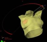
As modern radiation therapy evolves, new and novel techniques are proposed and/or established treatment modality is applied to new area.
Feasibility study, technical validations and clinical implementations are the subjects of many physics research. Quantitative evaluation of the new techniques against established standard methods are necessary to prove the clinical potential. Active areas of research in this group include but are not limited to the following:
Dynamic electron arc beam therapy with synchronized couch motion for tumors at shallow depth
Application of volumetric modulated radiation therapy (VMAT) to partial breast irradiation
Image-guided single fraction partial breast radiotherapy
Funding Sources
None
Selected Publications
Jian-Jian Qiu, Zheng Chang, Q. Jackie Wu, Sua Yoo, Janet Horton, Fang-Fang Yin, Impact of Volumetric Modulated Arc Therapy (v-MAT) Treatment Technique for Partial Breast Irradiation, International journal of radiation oncology, biology, physics 2010; 78:288-296.
Manisha Palta, Sua Yoo, Justus D. Adamson, Leonard R. Prosnitz, Janet K. Horton, Preoperative Single Fraction Partial Breast Radiotherapy for Early-Stage Breast Cancer, International journal of radiation oncology, biology, physics 2012; 82:37-42.
Investigator
Mark Oldham
Description
The lab pursues two main avenues of research. The first involves developing and applying new methods of high resolution 3D dosimetry. A range of uniquely capable state-of-the-art 3D dosimetry systems have been developed with funding support from the National Institute of Health. These systems are currently being applied to a diverse range of challenges in both the clinical (radiation therapy) and research domains. The second direction focuses on developing the new optical bio-imaging techniques of optical-computed-tomography (optical-CT), and optical-emission-computed-tomography (optical-ECT). These techniques have the potential to provide uniquely useful information on biological processes in bulk tumor and tissue samples.
Zheng (Jim) Chang, Jackie Wu
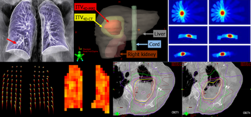
The management of respiratory motion in radiation oncology is one subject of important physics research. Respiratory motion affects all tumor sites in the thorax and abdomen.
Our research aims to improve the management of tumor motion via developing and evaluating novel 4D imaging techniques (4D-MRI, 4D-CBCT, 4D-PET, 4D-DTS) and 4D dose calculation methodologies, modeling and predicting lung and tumor respiratory motion, assessing treatment outcome of moving tumors, investigating correlations between internal tumor motion and external surrogate motion and clinically implementing new motion management techniques.
NIH - NCI Golfers Against Cancer (GAC) Foundation Philips Health System Varian Medical System
Cai J, Chang Z, O’Daniel J, Yoo S, Ge H, Kelsey C, Yin FF. Investigation of Sliced Body Area (SBA) as Respiratory Surrogate. J Am Clin Med Phys 2013;14(1):71-80.
Panta RK, Segars WP, Yin FF, Cai J. Implementing 4D-XCAT Phantom for 4D Radiotherapy Research. J Can Res Ther 2012;8:565-570.
Vergalasova I, Cai J, Yin FF. A novel technique for markerless, self-sorted 4D CBCT: feasibility study. Med Phys 2012 39(3):1442-1451.
Cai J, Chang Z, Wang Z, Segars WP, Yin FF. Four-dimensional Magnetic Resonance Imaging (4D-MRI) using Body Area as Internal Respiratory Surrogate: a Feasibility Study. Med. Phys 2011 38(12):6384-6394.
Wu QJ, Thongphiew D, Wang Z, Willett C, Marks L, Yin FF, Development of a 4D dosimetry simulation system in radiotherapy, Int. J. Biomedical Engineering and Technology, Vol. 8, Nos. 2/3, 201, 230-244 2012.
Jennifer O’Daniel, Zheng (Jim) Chang, Mark Oldham, Jackie Wu, Sua Yoo
Quality assurance (QA), both equipment-specific and patient-specific, is an essential component of safe radiotherapy treatments.
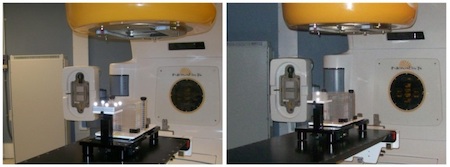
Our research team is focused on (1) improving the efficiency and effectiveness of current QA techniques and (2) developing new QA methods for novel technology. Current research projects include developing 3D QA tools, determining the correlation between traditional QA analysis criteria and the accuracy of patient treatment, quantifying the accuracy of novel radiotherapy devices (ex. flattening-filter free linear accelerator for breast radiotherapy), and creating methods for the QA of adaptive radiotherapy.
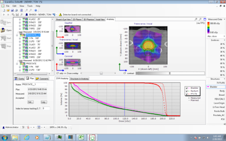
Scandidos, Inc.
Dube S, O’Daniel J, and Orton C. Point/Counterpoint: TG-142 is unwarranted for IGRT QA. Med Phys 2013; 40(1): 3987.
Chang Z, Bowsher J, Cai J, Yoo S, Wang S, Adamson J, Ren L, and Yin FF. Imaging system QA of a medical accelerator, Novalis Tx, for IGRT as per TG 142: Our 1 year experience. J Appl Clin Med Phys 2012; 13:113-140.
Oldham M, Thomas A, O’Daniel J, Juang T, Ibbott G, Adamovics J, and Kirkpatrick JP. A quality assurance method that utilitzes 3D dosimetry and facilitates clinical interpretation. Int J Radiat Oncol Biol Phys 2012; 84(2): 540-546.
O’Daniel J, Das S, Wu QJ, and Yin FF. Volumetric-Modulated Arc Therapy: Effective and Efficient End-to-End Patient-Specific Quality Assurance. Int J Radiat Oncol Biol Phys 2012; 84(5): 1567-74.
Chang Z, O’Daniel J, Yin FF. Quality assurance in adaptive radiation therapy, in Adaptive Radiation Therapy, edited by X. Allen Li. CRC Press, Taylor & Francis Group LLC, 2011.
Chang Z, Liu T, Cai J, Chen Q, Wang Z, and Yin FF. Evaluation of integrated respiratory gating systems on a Novalis Tx system. J Appl Clin Med Phys 2011; 12: 71-79.
Oana Craciunescu, Justus Adamson, Sheridan Meltsner, Mark Oldham
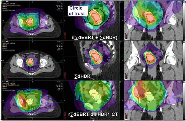
Image-guidance plays an important role in modern radiation therapy, predominantly in external beam planning and delivery. In recent years, with the advent of high/pulsed dose rate afterloading technology, advanced treatment planning systems, and CT and MRI compatible applicators, image-guided adaptive brachytherapy treatments (IGABT) are now achievable. With image guidance, the target can be delineated more precisely, resulting in delivering more controlled doses of radiation to the target while sparing surrounding healthy tissue.
Our group has been active in understand the benefits of IGARBT in general, MR-based in particular with emphasis on: applicator characterization, image registration, planning techniques in general, and the use of inverse planning in particular, role of model-based dose calculation algorithms, adaptive strategies, intrafraction variability, in-vivo dosimetry, dose summation with external beam treatments.
Radiation Oncology Department Support
R. McMahon, T. Zhuang, B. Steffey, H. Song, O. Craciunescu,” Commissioning of Varian Ring & Tandem HDR Applicators: Reproducibility and Inter-Observer Variability of Dwell Position Offsets”, Journal of Applied Medical Physics, vol 12, (4), 50-62, 2011.
Justus Adamson, Joseph Newton, Beverly Steffey, Jing Cai, Mark Oldham, Junzo Chino, and Oana Craciunescu, “Commissioning a CT compatible LDR tandem and ovoid applicator using 3D dosimetry, Medical Physics, 39, 4515-4523, 2012.
Pierquet M, Craciunescu O, Steffey B, Song H, Oldham M, “On the Feasibility of Verification of 3D Dosimetry Near Brachytherapy Sources Using PRESAGE/Optical-CT”, J. Phys.: Conf. Ser. 250 012091, 2010.
Chino J, Maurer J, Steffey B, Cai J, Adamson J, Craciunescu O, “IS AN MRI REQUIRED ON EACH FRACTION? AN EXPERIENCE WITH MRI GUIDED BRACHYTHERAPY FOR CERVICAL CANCER”, O. Radiotherapy and Oncology vol. 103 May, 2012. p. S107.
Craciunescu, J. Sánchez Mazón, L. Lan, J. Maurer, B. Steffey, J. Cai, J. Adamson, J. Chino, DOSIMETRIC IMPACT OF NOT CORRECTING FOR THE DISTAL SHIFT REPORTED IN VARIAN TANDEM AND RING (T&R) APPLICATORS”, J. Radiotherapy and Oncology vol. 103 May, 2012. p. S137
B. Steffey, O. Craciunescu, J. Cai, J. Adamson, J. Chino, “CLINICAL ASSESSMENT OF THE HDR CAPRI APPLICATOR”, Radiotherapy and Oncology vol. 103 May, 2012. p. S142.
J Adamson, J Newton, B Steffey, J Cai, J Adamovics, M Oldham, J Chino, and O Craciunescu, “Commissioning a CT Compatible LDR T&O Applicator Using Analytical Calculation with ID and 3D Dosimetry”, Med. Phys. 39, 3612 (2012).
J. Cai, J. Chino, Y. Qin, T.R. De Oliveira, J. Adamson, B. Steffey, O. Craciunescu, Feasibility of MR-alone-based Brachytherapy Treatment Planning Using a Titanium Tandem and Ring Applicator for Cervical Cancer, International Journal of Radiation Oncology*Biology*Physics, Volume 84, Issue 3, Supplement, 1 November 2012, Page S803.
Junzo Chino MD, Sheridan Meltsner PhD, Yun Yang PhD, Beverley Steffey MS, Jing Cai PhD, and Oana Craciunescu PhD, “Vaginal Dose in the Era of Image Guided Brachytherapy”, ABS 2013.
Oana Craciunescu PhD , Lei Ding, Jing Cai PhD, Beverley Steffey MS, Sheridan Meltsner PhD, Yun Yang PhD, Junzo Chino MD, “Intelligent Dose Summation from Multimodality Treatment of Cervical Cancer: A Case Study”, ABS 2013.
Y. Yang, S. Yan, J. Cai, B. Steffey, S. Meltsner, A. Thomas, F. Yin, O. Craciunescu, “Comprehensive Assessment Of Dose Variation Due to Ir-192 Source Position Within Varian Ring Using Monte Carlo Methods”, AAPM 2013.
Adria Vidovici et al, “Evaluation of a novel radiochromic dosimetry system for in-vivo dose verification in organs at risk in HDR intracavitary gynecological brachytherapy”, AAM 2013.
Research and Education Symposia
Craciunescu, O, Cai, J, Kirisits, C., de Leeuw, A., “Image Guided Adaptive Brachytherapy for Cervical Cancer”, AAPM, July 2012.
Craciunescu, O,Cai, J., Chino, J., “Implementing MR-Guided Adaptive Brachytherapy for Cervical Cancer”, AAPM 2013.
Jackie Wu, Qiuwen Wu
In current practice, IMRT/VMAT planning takes an experienced planner 1-6 hours, using an iterative, trial-and-error approach. Even with this effort the search for patient-specific optimal organ sparing is still strongly influenced by planner’s experience. Significant variations in plan quality have been observed at different institutions. Experienced centers are generally more capable of maximizing the dosimetric advantages of IMRT/VMAT. The knowledge and experience of an IMRT/VMAT treatment team is of great importance to realize the full benefits of this advanced technology.
This project focuses on developing knowledge models for guiding IMRTVMAT planning. The models are carefully developed from databases of high quality clinical plans, guidelines from clinical studies, as well as personal experience of expert planners. This comprehensive modeling approach with a strong focus on clinical practice is a distinguishing feature of this technology. These advanced models, will (1) capture human expert knowledge in designing high quality plans, (2) predict the optimal organ sparing that is specific to an individual patient, and (3) guide the design of the treatment plan to achieve optimal dose distribution with improved efficiency.
NIH - NCI Varian Medical System
Wu QJ, Li T, Wu Q, and Yin FF. Adaptive radiation therapy: technical components and clinical applications. Cancer J 17:182-9, 2011.
Li T, Thongphiew D, Zhu X, Lee WR, Vujaskovic Z, Yin FF, and Wu QJ. Adaptive prostate IGRT combining online re-optimization and re-positioning: a feasibility study. Phys. Med. Biol. 56: 1243, 2011.
Zhu X, Ge Y, Li T, Thongphiew D, Yin FF, and Wu QJ. A planning quality evaluation tool for prostate adaptive IMRT based on machine learning, Med. Phys. 38, 719: 723, 2011.
Li T, Zhu X, Thongphiew D, Lee WR, Vujaskovic Z, Wu Q, Yin FF, and Wu QJ. On-line Adaptive Radiation Therapy (ART): Feasibility And Clinical Study, J Oncol., vol. 2011.
Yuan L, Ge Y, Lee WR, Yin FF, Kirkpatrick JP, Wu QJ. Quantitative analysis of the factors which affect the interpatient organ-at-risk dose sparing variation in IMRT plans. Med Phys. 2012 Nov; 39(11):6868-78.
James Bowsher
Onboard imaging – as the patient is in position for treatment – is essential in radiation therapy. Currently onboard imaging is performed predominantly by cone-beam CT, which has limited capability for functional and molecular (F&M) imaging. Yet cancer is distinguished from surrounding healthy tissue largely by F&M characteristics. The purpose of this work is to develop single-photon emission computed tomography (SPECT) methods for F&M imaging onboard radiation therapy machines. These methods may also improve imaging for other tasks in which only a limited region of the full patient cross-section is of primary interest, such as in nuclear cardiology.
NIH National Cancer Institute
S Yan, J Bowsher, F Yin: Respiratory Sorted Imaging Using Region-Of-Interest Robotic Multi-Pinhole SPECT System. Presentation at the 55th Annual Meeting of the American Association of Physicists in Medicine, August 4-8, Indianapolis, IN, 2013. Medical Physics, 2013.
S Yan, J Bowsher, S Yoo, F Yin: On-Board Robotic Multi-Pinhole SPECT System for Prone Breast Imaging. Presentation at the 55th Annual Meeting of the American Association of Physicists in Medicine, August 4-8, Indianapolis, IN, 2013. Medical Physics, 2013.
J Bowsher, S Yan, F Yin: Robotic Multi-Pinhole Scenarios for SPECT Molecular and Functional Imaging Onboard and in Other Applications. Presentation at the 55th Annual Meeting of the American Association of Physicists in Medicine, August 4-8, Indianapolis, IN, 2013. Medical Physics, 2013.
S Yan, J Bowsher, F Yin: Functional and Molecular Imaging of the Axilla as the Patient Is in Position for Radiation Therapy Using a Robotic Multi-pinhole SPECT System. International Journal of Radiation Oncology Biology Physics 84(3) S247-S8, 2012.
S Yan, J Bowsher, W Giles, F Yin: A Line-Source Method for Aligning Onboard-Robotic-Pinhole and Other SPECT-Pinhole Systems. Presented at the 54th Annual Meeting of the American Association of Physicists in Medicine, July 29 - Aug 2, Charlotte, NC, 2012. Medical Physics 39(6) 4011, 2012.
J Bowsher, S Yan, J Roper, W Giles, F Yin: A Robotic Multi-Pinhole SPECT System for Onboard and Other Region-Of-Interest Imaging. Presented at the 54th Annual Meeting of the American Association of Physicists in Medicine, July 29 - Aug 2, Charlotte, NC, 2012. Medical Physics 39(6) 3887, 2012.
JE Bowsher, S Yan, JR Roper, WM Giles, F Yin: SPECT Imaging Onboard Radiation Therapy Machines. Invited Talk at SPIE Optics and Photonics: Optical Engineering and Applications: Medical Applications of Radiation Detectors, August 21-25, 2011, San Diego, CA.
Justus Adamson, William Giles
The radiosurgery research group is focused on improving treatment planning techniques and quality assurance methods for linear accelerator based radiosurgery.
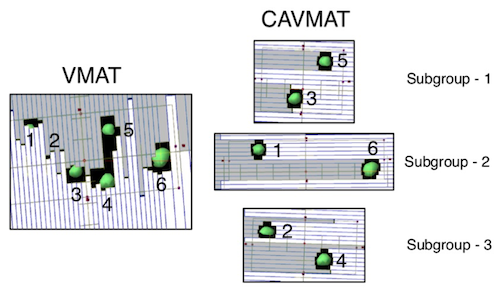
Conformal Arc Informed Volumetric Modulated Arc Therapy (CAVMAT) One current direction includes developing and refining a novel approach to stereotactic radiosurgery treatment planning for multiple brain metastases called Conformal Arc Informed VMAT (CAVMAT). When treating brain metastases there are several techniques that may be used, such as dynamic conformal arcs and VMAT. While effective and intuitive, dynamic conformal arcs suffer from a lack of modulation flexibility and extended treatment times. Conversely, VMAT is highly flexible and conformal, but may produce overly modulated and unintuitive MLC trajectories. In multi-target cases these unintuitive trajectories can lead to dose bridging between targets and irregular shaped isodose distributions.
CAVMAT is a hybrid technique, combining the intuitive MLC motions of dynamic conformal arc plans with the flexibility of VMAT inverse optimization. CAVMAT produces more intuitive dose distributions, reduces dose bridging and substantially spares healthy tissue without compromising plan quality.
Whereas VMAT may partially block targets or create unnecessary MLC openings, CAVMAT divides targets into subgroups, prioritizing effective collimation. The subgroups serve as a starting point for inverse optimization.
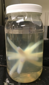
Fast and Comprehensive Verification of Radiation and Imaging Isocenter We are also working on developing a fast and comprehensive method to directly measure radiation isocenter uncertainty and coincidence with the kV-CBCT imaging system using 3D polymer gel dosimetry. We utilize novel N-isopropylacrylamide (NIPAM) gel dosimeters which have the unique characteristic of manifesting delivered dose as a change in density, and can thus be read out using CT and on-board CBCT. For comprehensive isocenter verification, a NIPAM dosimeter is irradiated at eight unique couch/gantry combinations, CBCT images are immediately acquired, radiation profile is detected per beam and the displacement from the imaging isocenter is quantified using MATLAB analysis. Setup, irradiation and CBCT readout can be performed within a typical QA slot.
Jim Chang, Justus Adamson
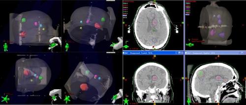
Stereotactic radiosurgery (SRS) requires treatment with high precision. It's always a challenge during SRS planning, localization and delivery to ensure the required high accuracy. This work aims to use advanced technologies such as image guidance and high definition MLC to develop techniques for precise and efficient SRS treatment.
Brainlab AG Varian Medical System
Z. Wang, J. Kirkpatrick, Z. Chang, J. O’Daniel, C. Willett, F-F. Yin. RapidArc for Treatment of Intracranial Multi-Focal Stereotactic Radiosurgery. ISRS 2009 Z. Wang, J. Nelson, S. Yoo, Q. J. Wu, J. Kirkpatrick, L. B. Marks, F-F Yin.
Refinement of Treatment set up and Target Localization Accuracy Using 3D Cone Beam CT for Stereotactic Body Radiation Therapy. Int. J. Radiat. Oncol. Biol. Phys. 73(2), p.571-7, (2009).
F-F. Yin, Z. Wang, S. Yoo, Q. J. Wu, J. Kirkpatrick, N. Larrier, J. Meyer, C. G. Willett, L. B. Marks, Integration of Cone-Beam CT in Stereotactic Body Radiation Therapy. Tech. Cancer Res. Treat. 7 (2), p.133-140, (2008).
Wang Z, Thomas A, Newton J, Ibbott G, Deasy J, Oldham M. Dose Verification of Stereotactic Radiosurgery Treatment for Trigeminal Neuralgia with Presage 3D Dosimetry System. J Phys Conf Ser. 2010 Dec 7;250(1). pii: 012058.
Z. Wang, Q. J. Wu, L. B. Marks, N. Larrier, and F-F. Yin. Cone-Beam CT Localization of Internal Target Volumes for Stereotactic Body Radiotherapy of Lung Lesions. Int. J. Radiat. Oncol. Biol. Phys. 69(5), p.1618-24, (2007).
Zheng Chang, Qiuwen Wu, Sua Yoo
Recent developments in image-guidance and immobilization enable target localization with increased accuracy, in order to deliver radiation more precisely to the tumor while sparing adjacent healthy tissue. With such improvements in imaging techniques, image guided radiation therapy (IGRT) has been widely adopted into clinical practice. Current active research projects includes 6 degree-of-freedom image guided SRS/SBRT, deformable registration for breast SBRT and 3D surface imaging for head and neck cancer radiotherapy.
Jinli Ma, Zheng Chang, Q. Jackie Wu , Zhiheng Wang, John P. Kirkpatrick, Fang-Fang Yin, ExacTrac X-ray 6 Degree-of-Freedom Image Guidance for Intracranial Noninvasive Stereotactic Radiotherapy: Comparison with kilo-voltage Cone-beam CT, Radiotherapy and Oncology 2009; 93:602–608.
Zheng Chang, Zhiheng Wang, Jinli Ma, Jennifer C. O’Daniel, John Kirkpatrick, and Fang-Fang Yin, 6D Image Guidance for Spinal Noninvasive Stereotactic Body Radiation Therapy: comparison between Exactrac X-ray 6D with Kilo-voltage Cone Beam CT, Radiotherapy and Oncology. 2010; 95: 116–121.
Olga Gopan, Qiuwen Wu, Evaluation of the accuracy of a 3D surface imaging system for patient setup in head and neck cancer radiotherapy.Int J Radiat Oncol Biol Phys. 2012;84(2):547-52.
Devon Godfrey, Zheng Chang, Jackie Wu, Sua Yoo, James Bowsher
IGRT has been widely applied in radiation therapy to use various imaging tools to improve the localization accuracy of the treatment. With the development of flat panel detectors, cone-beam CT (CBCT) has become a key imaging tool in IGRT, which is able to provide volumetric information about the patient for 3D or 4D target localization. MRI is another valuable tool under intensive study which is useful for target delineation and treatment assessment. Our group’s focus is to develop novel image acquisition and reconstruction techniques to improve the image quality and reduce imaging dose for various imaging modalities in IGRT.

Specifically we are focusing on the following directions:
- Imaging dose reduction using digital tomosynthesis (DTS). DTS only uses limited angle projections to reconstruct quasi-3D images; therefore it has much lower imaging dose than CBCT. This project is to evaluate the efficacy of DTS image guidance for different anatomic sites, including head-and-neck, prostate, breast, liver and lung.
- Development of clinical platform for DTS application. This project focuses on accelerating reconstruction using graphics card and developing user-friendly GUI interface for clinical use.
- Image reconstruction using prior knowledge and deformation models. A new method is developed to reconstruct full 3D images from limited-angle projections using patient prior knowledge and deformation models. Different deformation models, including PCA based motion models (MM) and free-form deformation (FD) model, are explored to improve the accuracy and efficiency.
- CBCT scatter correction. A synchronized moving grid (SMOG) system is being developed to correct for scatter, image lag and gantry flex of the flat panel detector based cone-beam CT (CBCT) system.
- Dual source CBCT. A dual source CBCT system has been built and its performance is being characterized. Virtual monochromatic (VM) and linearly mixed (LM) CBCTs are also developed to investigate their potential applications in metal artifact reduction and contrast enhancement in IGRT.
- Marker-less self-sorted 4D-CBCT. To develop an automatic projection sorting algorithm based on Fourier transformation of the projections.
- Fast reconstruction and processing of MR water-fat imaging, angiography, and quantitative functional imaging such as diffusion and perfusion imaging along with its clinical applications.
NIH-NCI Varian Medical System
D. J. Godfrey, F. F. Yin, M. Oldham, S. Yoo, C. Willett, “Digital tomosynthesis using an on-board kV imaging device,” Int. J. Radiat. Oncol. Biol. Phys. 65(1), 8-15 (2006).
Q. J. Wu, D. J. Godfrey, Z. Wang, J. Zhang, S. Zhou, S. You, D. Brizel, F. Yin, ”On-board patient positioning for head and neck IMRT: comparing digital tomosynthesis to kV radiography and cone-beam CT.” Int. J. Radiat. Oncol. Biol. Phys. 69(2), 598-606 (2007).
D. J. Godfrey, L. Ren, H. Yan, Q. Wu, S. Yoo, M. Oldham, F. Yin, “Evaluation of three types of reference image data for external beam radiotherapy target localization using digital tomosynthesis (DTS).” Med. Phys. 34(8) 3374-3384 (2007).
H. Yan, L. Ren, D. J. Godfrey, F. Yin, “Accelerating reconstruction of reference digital tomosynthesis using graphics hardware.” Med. Phys. 34(10), 3768-3776 (2007).
L. Ren, D. J. Godfrey, H. Yan, Q. J. Wu, F. Yin, “Automatic registration between reference and on-board digital tomosynthesis images for positioning verification.” Med. Phys. 35, 664 (2008).
H. Yan, D. J. Godfrey, F. Yin, “Fast reconstruction of digital tomosynthesis using on-board images.” Med. Phys. 35, 2162 (2008).
L. Ren, J. Zhang, D. Thongphiew, D. J. Godfrey, Z. Wang, F. Yin, “A novel digital tomosynthesis (DTS) reconstruction method using a deformation field map.” Med. Phys. 35, 3110 (2008).
J. Maurer, D. J. Godfrey, Z. Wang, F. Yin, “On-board four-dimensional digital tomosynthesis: First experimental results.” Med. Phys. 35, 3574 (2008).
S. Yoo, Q. J. Wu, D. J. Godfrey, H. Yan, L. Ren, S. Das, W. R. Lee, F. Yin, “Clinical evaluation of positioning verification using digital tomosynthesis (DTS) based on bony anatomy and soft tissues for prostate image-guided radiation therapy (IGRT).” Int. J. Radiat. Oncol. Biol. Phys. 73(1):296-305 (2009).
J. Zhang, Q. J. Wu, D. J. Godfrey, T. Fatunase, L. B. Marks, F. Yin, “Comparing digital tomosynthesis to cone-beam CT for position verification in patients undergoing partial breast irradiation,” Int. J. Radiat. Oncol. Biol. Phys. 73(3):952-957 (2009).
Z. Chang, Q. Xiang, J. Ji, and F.F. Yin, “Efficient Multiple Acquisitions by Skipped Phase Encoding and Edge Deghosting (SPEED) Using Shared Spatial Information,” Magn. Reson. Med. 61:229–233 (2009).
Z. Chang, Q. Xiang, H. Shen, and F.F. Yin, “Accelerating Non-Contrast-Enhanced MR Angiography with Inflow Inversion Recovery Imaging by Skipped Phase Encoding and Edge Deghosting (SPEED),” Journal of Magnetic Resonance Imaging 31:757-765 (2010).
J Maurer, T Pan, F Yin, “Slow gantry rotation acquisition technique for on-board four-dimensional tomosynthesis.” Med Phys 37:921-933 (2010).
J. Jin, L. Ren, Q. Liu, J. Kim, N. Wen, H. Guan, B. Movsas, I. Chetty, “Combining scatter reduction and correction to improve image quality in cone-beam computed tomography (CBCT)”, Med. Phys. 37, 5634-5644, (2010).
Q. J. Wu, J. Meyer, J. Fuller, D. Godfrey, Z. Wang, J. Zhang, F. Yin, “Digital tomosynthesis for respiratory gated liver treatment: Clinical feasibility for daily image guidance,” Int. J. Radiat. Oncol. Biol. Phys.79(1):289-296 (2011).
I. Vergalasova, J. Cai, F.F. Yin, “A novel technique for markerless, self-sorted 4D CBCT: feasibility study,” Med Phys 39(3):1442-1451 (2012).
L. Ren, I. Chetty, J. Zhang, J. Jin, Q.J. Wu, H. Yan, D.M. Brizel, W.R. Lee, B. Movsas, F. Yin, “Development and clinical evaluation of a three-dimensional cone-beam computed tomography estimation method using a deformation field map,” Int J Radiat Oncol Biol Phys, 82(5): 1584-93 (2012).
L. Ren, F. Yin, I. Chetty, D. Jaffray, and J. Jin, “Feasibility study of a synchronized-moving-grid (SMOG) system to improve image quality in Cone-Beam Computed Tomography (CBCT)”, Med. Phys., 39(8), 5099-5110, (2012).
H. Li, W. Giles, L. Ren, J. Bowsher and F.F. Yin, “Implementation of Dual-Energy Technique for Virtual Monochromatic and Linearly Mixed CBCTs”, Med. Phys. 39(10), 6056-64, 2012.
Z. Chang, Q. Xiang, H. Shen, J. Ji and F.F. Yin, "Accelerating Phase Contrast MR Angiography by Simplified Skipped Phase Encoding and Edge Deghosting with Array Coil Enhancement,” Med. Phys., 39:1247-1252 (2012).
H. Li, W. Giles, J. Bowsher and F.F. Yin, “A Dual Cone-Beam CT System for Image Guided Radiotherapy: Initial Performance Characterization”, Med. Phys. 40(2), 021912, 2013.
- Search by keyword
- Search by citation
Page 1 of 2
Microwave open-ended coaxial dielectric probe: interpretation of the sensing volume re-visited
Tissue dielectric properties are specific to physiological changes and consequently have been pursued as imaging biomarkers of cancer and other pathological disorders. However, a recent study (Phys Med Biol 52...
- View Full Text
Diagnosis of cervical cells based on fractal and Euclidian geometrical measurements: Intrinsic Geometric Cellular Organization
Fractal geometry has been the basis for the development of a diagnosis of preneoplastic and neoplastic cells that clears up the undetermination of the atypical squamous cells of undetermined significance (ASCUS).
Reviewer acknowledgement 2013
The editors of BMC Medical Physics would like to thank all our reviewers who have contributed to the journal in Volume 13 (2013).
Validity of actigraphs uniaxial and triaxial accelerometers for assessment of physical activity in adults in laboratory conditions
Few studies to date have directly compared the Actigraphs GT1M and the GT3X, it would be of tremendous value to know if these accelerometers give similar information about intensities of PA. Knowing if output ...
Real-time prostate motion assessment: image-guidance and the temporal dependence of intra-fraction motion
The rapid adoption of image-guidance in prostate intensity-modulated radiotherapy (IMRT) results in longer treatment times, which may result in larger intrafraction motion, thereby negating the advantage of im...
Predictions of CD4 lymphocytes’ count in HIV patients from complete blood count
HIV diagnosis, prognostic and treatment requires T CD4 lymphocytes’ number from flow cytometry, an expensive technique often not available to people in developing countries. The aim of this work is to apply a ...
Dose mapping sensitivity to deformable registration uncertainties in fractionated radiotherapy – applied to prostate proton treatments
Calculation of accumulated dose in fractionated radiotherapy based on spatial mapping of the dose points generally requires deformable image registration (DIR). The accuracy of the accumulated dose thus depend...
The 2D Hotelling filter - a quantitative noise-reducing principal-component filter for dynamic PET data, with applications in patient dose reduction
In this paper we apply the principal-component analysis filter (Hotelling filter) to reduce noise from dynamic positron-emission tomography (PET) patient data, for a number of different radio-tracer molecules....
Multiscale forward electromagnetic model of uterine contractions during pregnancy
Analyzing and monitoring uterine contractions during pregnancy is relevant to the field of reproductive health assessment. Its clinical importance is grounded in the need to reliably predict the onset of labor...
The study of radiosensitivity in left handed compared to right handed healthy women
Radiosensitivity is an inheriting trait that mainly depends on genetic factors. it is well known in similar dose of ionizing radiation and identical biological characteristics 9–10 percent of normal population...
Comparison of the dosimetries of 3-dimensions Radiotherapy (3D-RT) with linear accelerator and intensity modulated radiotherapy (IMRT) with helical tomotherapy in children irradiated for neuroblastoma
Intensity modulated radiotherapy is an efficient radiotherapy technique to increase dose in target volumes and decrease irradiation dose in organs at risk. This last objective is mainly relevant in children. H...
Navigator channel adaptation to reconstruct three dimensional heart volumes from two dimensional radiotherapy planning data
Biologically-based models that utilize 3D radiation dosimetry data to estimate the risk of late cardiac effects could have significant utility for planning radiotherapy in young patients. A major challenge ari...
Preclinical multimodality phantom design for quality assurance of tumor size measurement
Evaluation of changes in tumor size from images acquired by ultrasound (US), computed tomography (CT) or magnetic resonance imaging (MRI) is a common measure of cancer chemotherapy efficacy. Tumor size measure...
Theoretical generalization of normal and sick coronary arteries with fractal dimensions and the arterial intrinsic mathematical harmony
Fractal geometry is employ to characterize the irregular objects and had been used in experimental and clinic applications. Starting from a previous work, here we made a theoretical research based on a geometr...
Differential radio-sensitivities of human chromosomes 1 and 2 in one donor in interphase- and metaphase-spreads after 60 Co γ-irradiation
Radiation-induced chromosome aberrations lead to a plethora of detrimental effects at cellular level. Chromosome aberrations provide broad spectrum of information ranging from probability of malignant transfor...
Prognostic implication of late gadolinium enhancement on cardiac MRI in light chain (AL) amyloidosis on long term follow up
Light chain amyloidosis (AL) is a rare plasma cell dyscrasia associated with poor survival especially in the setting of heart failure. Late gadolinium enhancement (LGE) on cardiac MRI was recently found to cor...
Average arterial input function for quantitative dynamic contrast enhanced magnetic resonance imaging of neck nodal metastases
The present study determines the feasibility of generating an average arterial input function (Avg-AIF) from a limited population of patients with neck nodal metastases to be used for pharmacokinetic modeling ...
Bone turnover markers are correlated with total skeletal uptake of 99m Tc-methylene diphosphonate ( 99m Tc-MDP)
Skeletal uptake of 99m Tc labelled methylene diphosphonate ( 99m Tc-MDP) is used for producing images of pathological bone uptake due to its incorporation to the sites of active bone turnover. This study was done to...
Repeatability of regional myocardial blood flow calculation in 82 Rb PET imaging
We evaluated the repeatability of the calculation of myocardial blood flow (MBF) at rest and pharmacological stress, and calculated the coronary flow reserve (CFR) utilizing 82 Rb PET imaging. The aim of the resea...
Chemotherapeutic treatment efficacy and sensitivity are increased by adjuvant alternating electric fields (TTFields)
The present study explores the efficacy and toxicity of combining a new, non-toxic, cancer treatment modality, termed Tumor Treating Fields (TTFields), with chemotherapeutic treatment in-vitro, in-vivo and in ...
Multiple window spatial registration error of a gamma camera: 133 Ba point source as a replacement of the NEMA procedure
The accuracy of multiple window spatial resolution characterises the performance of a gamma camera for dual isotope imaging. In the present study we investigate an alternative method to the standard NEMA proce...
Influence of increased target dose inhomogeneity on margins for breathing motion compensation in conformal stereotactic body radiotherapy
Breathing motion should be considered for stereotactic body radiotherapy (SBRT) of lung tumors. Four-dimensional computer tomography (4D-CT) offers detailed information of tumor motion. The aim of this work is...
Metabolism of no-carrier-added 2-[ 18 F]fluoro-L-tyrosine in rats
Several fluorine-18 labelled fluoroamino acids have been evaluated as tracers for the quantitative assessment of cerebral protein synthesis in vivo by positron emission tomography (PET). Among these, 2-[ 18 F]fluor...
NEO adjuvant therapy monitoring with P ET and CT in E sophageal C ancer (NEOPEC-trial)
Surgical resection is the preferred treatment of potentially curable esophageal cancer. To improve long term patient outcome, many institutes apply neoadjuvant chemoradiotherapy. In a large proportion of patie...
A computerized Infusion Pump for control of tissue tracer concentration during Positron Emission Tomography in vivo Pharmacokinetic/Pharmacodynamic measurements
A computer controlled infusion pump (UIPump) for regulation of target tissue concentration of radioactive compounds was developed for use in biological research and tracer development for PET.
Perfusion scanning using 99m Tc-HMPAO detects early cerebrovascular changes in the diabetic rat
99m Tc-HMPAO is a well-established isotope useful in the detection of regional cerebral blood flow. Diabetes gives rise to arterial atherosclerotic changes that can lead to significant end organ dysfunction, prom...
Computer-assisted lateralization of unilateral temporal lobe epilepsy using Z-score parametric F-18 FDG PET images
To evaluate the use of unbiased computer-assisted lateralization of temporal lobe epilepsy (TLE) by z-score parametric PET imaging (ZPET).
Comparison of 2D, 3D high dose and 3D low dose gated myocardial 82 Rb PET imaging
We compared 2D, 3D high dose (HD) and 3D low dose (LD) gated myocardial Rb-82 PET imaging in 16 normal human studies. The main goal in the paper is to evaluate whether the images obtained by a 3D LD studies ar...
Toxicology evaluation of radiotracer doses of 3'-deoxy-3'-[ 18 F]fluorothymidine ( 18 F-FLT) for human PET imaging: Laboratory analysis of serial blood samples and comparison to previously investigated therapeutic FLT doses
18 F-FLT is a novel PET radiotracer which has demonstrated a strong potential utility for imaging cellular proliferation in human tumors in vivo. To facilitate future regulatory approval of 18 F-FLT for clinical u...
Comparison of manual and semi-automated delineation of regions of interest for radioligand PET imaging analysis
As imaging centers produce higher resolution research scans, the number of man-hours required to process regional data has become a major concern. Comparison of automated vs. manual methodology has not been re...
Variation in heart rate influences the assessment of transient ischemic dilation in myocardial perfusion scintigraphy
Transient arrhythmias can affect transient ischemic dilation (TID) ratios. This study was initiated to evaluate the frequency and effect of normal heart rate change on TID measures in routine clinical practice.
Comparison of 18 F SPECT with PET in myocardial imaging: A realistic thorax-cardiac phantom study
Positron emission tomography (PET) imaging with fluorine-18 ( 18 F) Fluorodeoxyglucose (FDG) and flow tracer such as Rubidium-82 ( 82 Rb) is an established method for evaluating an ischemic but viable myocardium. How...
A statistical investigation of normal regional intra-subject heterogeneity of brain metabolism and perfusion by F-18 FDG and O-15 H 2 O PET imaging
The definite evaluation of the regional cerebral heterogeneity using perfusion and metabolism by a single modality of PET imaging has not been well addressed. Thus a statistical analysis of voxel variables fro...
Non-invasive imaging of atherosclerotic plaque macrophage in a rabbit model with F-18 FDG PET: a histopathological correlation
Coronary atherosclerosis and its thrombotic complications are the major cause of mortality and morbidity throughout the industrialized world. Thrombosis on disrupted atherosclerotic plaques plays a key role in...
Prognostic utility of sestamibi lung uptake does not require adjustment for stress-related variables: A retrospective cohort study
Increased 99m Tc-sestamibi stress lung-to-heart ratio (sLHR) has been shown to predict cardiac outcomes similar to pulmonary uptake of thallium. Peak heart rate and use of pharmacologic stress affect the interpret...
Frequency and severity of myocardial perfusion abnormalities using Tc-99m MIBI SPECT in cardiac syndrome X
Cardiac syndrome X is defined by a typical angina pectoris with normal or near normal (stenosis <40%) coronary angiogram with or without electrocardiogram (ECG) change or atypical angina pectoris with normal o...
Relationship of 99m technetium labelled macroaggregated albumin ( 99m Tc-MAA) uptake by colorectal liver metastases to response following Selective Internal Radiation Therapy (SIRT)
SIRT is an emerging treatment for liver tumours which relies on the selective uptake by tumour of 90 Y microspheres following hepatic arterial injection. Response rates of around 90% are reported. Hepatic arterial...
A preliminary study of neuroSPECT evaluation of patients with post-traumatic smell impairment
Most olfactory testings are subjective and since they depend upon the patients' response, they are prone to false positive results. The aim of this study was to use quantitative brain perfusion SPECT in order ...
Hydrophilic and lipophilic radiopharmaceuticals as tracers in pharmaceutical development: In vitro – In vivo studies
Scintigraphic studies have been performed to assess the release, both in vitro and in vivo , of radiotracers from tablet formulations. Four different tracers with differing physicochemical characteristics have bee...
Planar Tc99m – sestamibi scintimammography should be considered cautiously in the axillary evaluation of breast cancer protocols: Results of an international multicenter trial
Lymph node status is the most important prognostic indicator in breast cancer in recently diagnosed primary lesion. As a part of an interregional protocol using scintimammography with Tc99m compounds, the valu...
Use of segmented CT transmission map to avoid metal artifacts in PET images by a PET-CT device
Attenuation correction is generally used to PET images to achieve count rate values independent from tissue densities. The goal of this study was to provide a qualitative comparison of attenuation corrected PE...
Comparison of SPECT bone scintigraphy with MRI for diagnosis of meniscal tears
Scintigraphy has been considered as competitive to MRI, but limited data are available on the accuracy of single photon emission tomography (SPECT) compared with MRI for the assessment of meniscal tears. Our o...
Sodium pertechnetate (Na99mTcO 4 ) biodistribution in mice exposed to cigarette smoke
The biological effects of cigarette smoke are not fully known. To improve our understanding of the action of various chemical agents, we investigated the biodistribution of sodium pertechnetate (Na 99m TcO 4 ) in mic...
Evaluation of the clinical value of bone metabolic parameters for the screening of osseous metastases compared to bone scintigraphy
Bone metastases are common in many types of cancer. As screening methods different imaging modalities are available. A new approach for the screening of osseous metastases represents the measurement of bone me...
The hazards of lack of co-registration of ictal brain SPECT with MRI: A case report of sinusitis mimicking a brainstem seizure focus
Single photon emission computed tomography (SPECT) following injection of radiotracer during a seizure is known as ictal SPECT. Comparison of an ictal SPECT study to a baseline or interictal study can aid iden...
188 Re radiopharmaceuticals for radiosynovectomy: evaluation and comparison of tin colloid, hydroxyapatite and tin-ferric hydroxide macroaggregates
Radiosynovectomy is a therapy used to relieve pain and inflammation from rheumatoid arthritis and related diseases. In this study three 188 Re particulate compounds were characterized according to their physico-ch...
Comparison of Technetium-99m-MIBI imaging with MRI for detection of spine involvement in patients with multiple myeloma
Recently, radiopharmaceutical scanning with Tc-99m-MIBI was reported to depict areas with active bone disease in multiple myeloma (MM) with both high sensitivity and specificity. This observation was explained...
Assessment of diffuse Lewy body disease by 2-[18F]fluoro-2-deoxy-D-glucose positron emission tomography (FDG PET)
Lewy body disease is, after Alzheimer's disease, the second most common cause of senile degenerative dementia with progressive cognitive deterioration, fluctuation of cognitive and motoric functions and psycho...
Physico-chemical characterisation and biological evaluation of 188-Rhenium colloids for radiosynovectomy
Radiosynovectomy is a type of radiotherapy used to relieve pain and inflammation from rheumatoid arthritis. In this study, 188-Rhenium ( 188 Re) colloids were characterized by physical and biological methodologies....
Re-HEDP : pharmacokinetic characterization, clinical and dosimetric evaluation in osseous metastatic patients with two levels of radiopharmaceutical dose
A study for pain relief therapy with 188 Re-HEDP was done in patients with bone metastases secondary to breast and prostate cancer.
BMC Medical Physics
ISSN: 1756-6649
Thank you for visiting nature.com. You are using a browser version with limited support for CSS. To obtain the best experience, we recommend you use a more up to date browser (or turn off compatibility mode in Internet Explorer). In the meantime, to ensure continued support, we are displaying the site without styles and JavaScript.
- View all journals
- Explore content
- About the journal
- Publish with us
- Sign up for alerts
Collection 04 September 2019
Medical physics
Medical physics is a very well-established field where advances tend to be of technical nature. however, physicists from other areas of physics can make unexpected contributions. new ideas, technology transfer and interdisciplinary collaborations can lead to exciting developments. this collection gathers various news, review and opinion pieces highlighting and discussing such trends..

Positronium in medicine and biology
In positron emission tomography, up to 40% of positron annihilation occurs through the production of positronium atoms in the patient’s body, whose decay could provide information about disease progression. New research is needed to take full advantage of this information.
- Paweł Moskal
- Bożena Jasińska
- Steven D. Bass

A sustainable future for nuclear imaging
Each year millions of patients benefit from diagnostic services enabled by advances in medical imaging. However, some services rely on the supply of technetium-99m from an ageing nuclear infrastructure. Kevin Charlton discusses new technologies to secure a sustainable supply.
- Kevin Charlton
Medical isotope production with the IsoDAR cyclotron
Jose R. Alonso and colleagues describe technical advances that will allow the proposed IsoDAR (isotope decay at rest) cyclotron — being developed for neutrino physics research — to produce many medical isotopes more efficiently than existing cyclotrons can.
- Jose R. Alonso
- Roger Barlow
- Loyd Hoyt Waites

Nucleation, mapping and control of cavitation for drug delivery
This Review describes how acoustic cavitation can be used to improve the delivery of drugs for the treatment of diseases such as cancer and stroke. Methods for seeding cavitation, treatment monitoring, and current and future clinical applications are described.
- Eleanor Stride
- Constantin Coussios
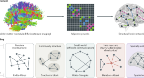
The physics of brain network structure, function and control
The brain is the quintessential complex system, boasting incredible feats of cognition and supporting a wide range of behaviours. Physics has much to offer in the quest to distil the brain’s complexity to a number of cogent organizing principles.
- Christopher W. Lynn
- Danielle S. Bassett
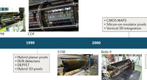
Applications of silicon strip and pixel-based particle tracking detectors
Advances in semiconductor technologies have enabled the development of numerous designs of silicon tracking detectors in particle physics. This Technical Review outlines the current state-of-the-art technologies and discusses challenges, future directions and some of the recent applications outside particle physics.
- Philip Allport
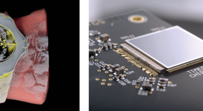
X-ray images in full colour
Detector technologies developed at CERN can produce stunning colour X-ray computed tomography images, but bringing them to hospitals is challenging.
- Zoe Budrikis
New physics for medical physics
This month we examine examples of how advances from different areas of physics can lead to medical applications.
Quick links
- Explore articles by subject
- Guide to authors
- Editorial policies
Health Physics
- Subscribe to journal Subscribe
- Get new issue alerts Get alerts
Secondary Logo
Journal logo, featured video.
Flash Player 9.0.0 is required for this Video. Get Adobe Flash Player .
Special Issue of the Global Conference of Radiation Topics (ConRad), Munich 2017
ConRad 2017, the Global Conference on Radiation Topics, Preparedness, Response, Protection and Research offered a unique opportunity for professional and multidisciplinary exchange of experience and expertise in the diversified field of radiation sciences. Within this special issue we are proud to present selected manuscript of our meeting.
ConRad takes place every 2 years in Munich under the auspices of the Bundeswehr Institute of Radiobiology. For more information about our next meeting check: www.radiation-medicine.de
Editor's Note: Getting Ready for the Move
Current supplement.

Published April 2022
Colleague's E-mail is Invalid
Your message has been successfully sent to your colleague.
Save my selection
Current Issue Highlights

Bisphosphonate Liposomes for Cobalt and Strontium Decorporation?
Health Physics. 127(4):463-475, October 2024.
- Abstract Abstract
- Permissions
- OPERATIONAL TOPICS


A Compendium of Radiation Safety Practices That Can Complement Organizational Worker Well-being Initiatives
Health Physics. 127(4):539-542, October 2024.
- Radiation Protection Abstracts
Health Physics. 127(4):550-553, October 2024.
- Health Physics Society Prospectus
THE HEALTH PHYSICS SOCIETY: An Affiliate of the International Radiation Protection Association (IRPA)
Health Physics. 127(4):554, October 2024.
- Health Physics Society Affiliate Members
HEALTH PHYSICS SOCIETY . 2024 AFFILIATE MEMBERS
Health Physics. 127(4):555, October 2024.
Current Issue
Absolute method for measuring environmental radioactive materials using imaging plates.
Health Physics. 127(4):476-480, October 2024.
Research on Detection Efficiency of Imaging Plates for Alpha Particles Using Two Types of Imaging Plate
Health Physics. 127(4):481-489, October 2024.
Decontamination of Actinide-contaminated Injured Skin with Ca-DTPA Products Using an Ex Vivo Rat Skin Model
Health Physics. 127(4):490-503, October 2024.
Go to Full Text of this Article
Comparison of MCNP and Microshield Dose Savings Determinations for Remote Methods of Transuranic Contamination Characterization
Health Physics. 127(4):504-512, October 2024.
Investigating the Effects of Tube Current and Tube Voltage on Patient Dose in Computed Tomography Examinations with Principial Component Analysis and Cluster Analysis: Phantom Study
Health Physics. 127(4):513-519, October 2024.
Uranium Body Clearance Kinetics—A Long-term Follow-up Study of Retired Nuclear Fuel Workers
Health Physics. 127(4):520-535, October 2024.
Evaluation of Novel General Education Courses on Radiation Protection for Undergraduates
Health Physics. 127(4):543-548, October 2024.

- What is Medical Physics
- Our History
- Life at Duke
- News & Events
- About the Program
- Degree Requirements
- List of Courses
- Stats on Admission
- Admissions Prerequisites
- Financial Aid
- How to Apply
- Student Publications
Research Interests
- Facilities & Labs
- Full Faculty
- Associate, Adjunct, Educational Affiliate, & Emeritus
- Current Students
- Our Community
- Diversity, Equity & Inclusion
Four Tracks, One Program
Our faculty are drawn from 5 departments: Radiology, Radiation Oncology, Radiation Safety, Biomedical Engineering, and Physics. In addition, each faculty affiliates with one or more of the program’s four tracks.
The tracks are shown below with representative topics of research highlighting just some of the strengths of our program.
Diagnostic Imaging (DI)
- advanced ultrasound imaging
- artificial intelligence for diagnosis, reconstruction, etc.
- dedicated breast CT
- detector and display characterization
- dual energy CT
- functional brain MRI
- molecular imaging
- MR microscopy
- MRI with hyperpolarized gas & fluorinated compounds
- neutron stimulated emission computed tomography
- optical interference tomography
- x-ray digital tomosynthesis
Radiation Therapy (RT)
- hyperthermia
- treatment planning optimization
- intensity modulated radiation therapy
- 3-D dosimetry
- oncological imaging and image analysis
- image guided radiation therapy
- stereotactic radiosurgery
- biological modelling of tumor and normal tissue dose responses
- intra-operative radiation therapy
Medical Health Physics (MHP)
- bone marrow dosimetry
- CT dosimetry: dual energy, hefnium filter, etc.
- energy modulated CBCT dosimetry
- Monte Carlo simulation in small animal and humans
- nano-fiber optic dosimetry: brachytherapy and other applications
- neutron environmental monitoring with TLDs
- parathyroid imaging: 4D CT vs. Sestamibi
- pediatric dosimetry: cath lab and head CT
Nuclear Medicine (NM)
- statistical iterative reconstruction algorithms
- whole body PET-CT fusion imaging
- dedicated PET and SPECT breast imaging systems
- novel ECT acquisition geometries
| is powered by WordPress. Read the Sites@Duke Express and , or . |

- Public & Media
- International
- Medical Physicist
- Quality & Safety
- Government Affairs
- Medical Physics Journal
- Journal of Applied Clinical Medical Physics
- WPSC Newsletter
- Research Spotlight
- Physics Today
- CT Protocols
- Medical Physics Practice Guidelines
- ACR-AAPM Practice Parameters and Technical Standards
- Annals of the ICRP
- Online ICRU Publications
- Online NCRP Publications
- Medical Physics Electronic Content (MPEC)
- Author Archives
- Surveys (Professional & Research)
- Copyright and Permissions
- Career Services
- Corporate Affiliates
- Links of Interest
- Advertising Opportunities

AAPM supports its mission by disseminating scientific and technical information in the discipline of medical physics.
| ( ) is our flagship publication with authors and subscribers throughout the world. It is available in print and online through individual and library subscriptions. Subscription provides online access and sophisticated search capability through the entire journal archive at no additional charge, and a significant number of featured articles are also available without charge. RSS feeds from Medical Physics provide notification of new articles published in selected topical areas regardless of subscription. | The (JACMP) is an open-access journal, which publishes papers that will help clinical medical physicists perform their responsibilities more effectively and efficiently for the increased benefit of the patient. |
| are technical reports on specific topics that are prepared by experts drawn from the AAPM membership who work together in Task Groups under the guidance of the AAPM Councils and Committees. All AAPM reports are published on-line with open-access without subscription. are reports created by AAPM Ad Hoc Committees that answer their specific charge. AAPM Monographs and Proceedings are reviews of medical physics topics and are primarily the output of many of the AAPM Summer Schools. These are available for purchase from the . from the AAPM Annual Meetings, recent AAPM Summer Schools other meetings are . are intended to provide the medical community with a clear description of the minimum level of medical physics support that AAPM would consider to be prudent in all clinical practice settings. Support includes but is not limited to staffing, equipment, machine access, and training. Originally commissioned by the Ad Hoc Committee on External Communications and Social Media, assists health professionals in explaining the benefits and risks of medical imaging to care providers, patients, family members, and the public. ACR-AAPM Practice Parameters and Technical Standards are the result of the collaboration of members of AAPM, ACR, and sometimes other organizations. Each has been reviewed by the membership of the contributing organizations and is subject to regular revisiting and updating as needed. | (High Dose per Fraction, Hypofractionated Treatment Effects in the Clinic) is the project of the AAPM Working Group on Biological Effects of Hypofractionated Radiotherapy/SBRT (WGSBRT). The WG includes physicists, radiation oncologists and radiation biologists and is charged with generating reports based on critical review of published data regarding both tumor and normal tissue response to the strongly hypofractionated regimens used in SBRT (SABR) treatments with suggestions for outcomes reporting standards that will facilitate future analyses. The project also includes several more speculative "Vision Papers" on the radiobiology of these treatments. The work is the result of the diligent efforts of numerous investigators, authors, reviewers and support personnel. generated by and approved by any AAPM committee with the intent of making such content available via the AAPM website. Miscellaneous Reports2021 Revision. . N. Ozturk, S.G. Armato III, M.L. Giger, C.F. Serago, and L.F. Ross. ; 40(4): 047001-1 - 047001-8. . Gray, JE, Archer, BR, Butler, PF, et al. 2005;235: 354-358. Report of AAPM Task Group No. 7 (Reference Values for Diagnostic X-Ray Examinations) of the Radiation Protection Committee.
|
Newsletters

Organizational Subscriptions
Please be mindful that the agreements that grant AAPM Members free access to these documents prohibit distribution beyond AAPM Members. Posting these documents online is a violation the agreements and could jeopardize AAPM's ability to retain this AAPM Member benefit in the future.

This Website publishes information of interest to members and beyond and acts as the portal to all AAPM publications and to the diverse activities of this Association. RSS feeds from the home page provide notification of new topical postings.
AAPM's Privacy Policy Use of the site constitutes your acceptance to its terms and conditions .
AAPM is a scientific, educational, and professional nonprofit organization devoted to the discipline of physics in medicine. The information provided in this website is offered for the benefit of its members and the general public, however, AAPM does not independently verify or substantiate the information provided on other websites that may be linked to this site.
Information
- Author Services
Initiatives
You are accessing a machine-readable page. In order to be human-readable, please install an RSS reader.
All articles published by MDPI are made immediately available worldwide under an open access license. No special permission is required to reuse all or part of the article published by MDPI, including figures and tables. For articles published under an open access Creative Common CC BY license, any part of the article may be reused without permission provided that the original article is clearly cited. For more information, please refer to https://www.mdpi.com/openaccess .
Feature papers represent the most advanced research with significant potential for high impact in the field. A Feature Paper should be a substantial original Article that involves several techniques or approaches, provides an outlook for future research directions and describes possible research applications.
Feature papers are submitted upon individual invitation or recommendation by the scientific editors and must receive positive feedback from the reviewers.
Editor’s Choice articles are based on recommendations by the scientific editors of MDPI journals from around the world. Editors select a small number of articles recently published in the journal that they believe will be particularly interesting to readers, or important in the respective research area. The aim is to provide a snapshot of some of the most exciting work published in the various research areas of the journal.
Original Submission Date Received: .
- Active Journals
- Find a Journal
- Proceedings Series
- For Authors
- For Reviewers
- For Editors
- For Librarians
- For Publishers
- For Societies
- For Conference Organizers
- Open Access Policy
- Institutional Open Access Program
- Special Issues Guidelines
- Editorial Process
- Research and Publication Ethics
- Article Processing Charges
- Testimonials
- Preprints.org
- SciProfiles
- Encyclopedia

Journal Menu
- Applied Sciences Home
- Aims & Scope
- Editorial Board
- Reviewer Board
- Topical Advisory Panel
- Instructions for Authors
- Special Issues
- Sections & Collections
- Article Processing Charge
- Indexing & Archiving
- Editor’s Choice Articles
- Most Cited & Viewed
- Journal Statistics
- Journal History
- Journal Awards
- Society Collaborations
- Conferences
- Editorial Office
Journal Browser
- arrow_forward_ios Forthcoming issue arrow_forward_ios Current issue
- Vol. 14 (2024)
- Vol. 13 (2023)
- Vol. 12 (2022)
- Vol. 11 (2021)
- Vol. 10 (2020)
- Vol. 9 (2019)
- Vol. 8 (2018)
- Vol. 7 (2017)
- Vol. 6 (2016)
- Vol. 5 (2015)
- Vol. 4 (2014)
- Vol. 3 (2013)
- Vol. 2 (2012)
- Vol. 1 (2011)
Find support for a specific problem in the support section of our website.
Please let us know what you think of our products and services.
Visit our dedicated information section to learn more about MDPI.
Medical Physics: Latest Advances and Prospects
- Print Special Issue Flyer
- Special Issue Editors
Special Issue Information
Benefits of publishing in a special issue.
- Published Papers
A special issue of Applied Sciences (ISSN 2076-3417). This special issue belongs to the section " Applied Physics General ".
Deadline for manuscript submissions: 20 January 2025 | Viewed by 19225
Share This Special Issue
Special issue editor.

Dear Colleagues,
The principles and methods of applied physics have long been applied to Medicine for the design of diagnostic and therapeutic techniques through the use of ionizing and non-ionizing radiation. Medical Physics covers all areas of applied physics research dealing with the prevention, diagnosis, and treatment of human diseases. Medical Physics encompasses both experimental and theoretical research and strongly involves computing. This Special Issue is dedicated to new developments in the field of Medical Physics, which includes (but is not limited to) radiation therapy, radiation protection, biomedical imaging, and related topics in health physics and biophysics, including space applications. Topics focusing on theoretical, computational, and experimental approaches to Medical Physics are welcome.
Dr. Ioanna Kyriakou Guest Editor
Manuscripts should be submitted online at www.mdpi.com by registering and logging in to this website . Once you are registered, click here to go to the submission form . Manuscripts can be submitted until the deadline. All submissions that pass pre-check are peer-reviewed. Accepted papers will be published continuously in the journal (as soon as accepted) and will be listed together on the special issue website. Research articles, review articles as well as short communications are invited. For planned papers, a title and short abstract (about 100 words) can be sent to the Editorial Office for announcement on this website.
Submitted manuscripts should not have been published previously, nor be under consideration for publication elsewhere (except conference proceedings papers). All manuscripts are thoroughly refereed through a single-blind peer-review process. A guide for authors and other relevant information for submission of manuscripts is available on the Instructions for Authors page. Applied Sciences is an international peer-reviewed open access semimonthly journal published by MDPI.
Please visit the Instructions for Authors page before submitting a manuscript. The Article Processing Charge (APC) for publication in this open access journal is 2400 CHF (Swiss Francs). Submitted papers should be well formatted and use good English. Authors may use MDPI's English editing service prior to publication or during author revisions.
- external beam radiotherapy
- brachytherapy
- radiopharmaceutical therapy
- biomedical imaging
- radiation protection
- radiation biophysics
- medical biophysics
- radiation dosimetry
- radiation transport
- Monte Carlo simulations
- space radiation health
- Ease of navigation: Grouping papers by topic helps scholars navigate broad scope journals more efficiently.
- Greater discoverability: Special Issues support the reach and impact of scientific research. Articles in Special Issues are more discoverable and cited more frequently.
- Expansion of research network: Special Issues facilitate connections among authors, fostering scientific collaborations.
- External promotion: Articles in Special Issues are often promoted through the journal's social media, increasing their visibility.
- e-Book format: Special Issues with more than 10 articles can be published as dedicated e-books, ensuring wide and rapid dissemination.
Further information on MDPI's Special Issue polices can be found here .
Published Papers (10 papers)
Jump to: Review

Jump to: Research

Further Information
Mdpi initiatives, follow mdpi.

Subscribe to receive issue release notifications and newsletters from MDPI journals

- AAPM Members
- Library Sales
- Copying and Other Use
- Other AAPM Publications
- Deputy Editors
- Editorial Board
- AE Guidelines
- Impact Factor 4.506
- Instructions for Authors
- Submit an Article
- Editorial Policies
- Revision Instructions
- Preparing Your Article Files for Publication
- Login to the System
- Submitting Supporting Material
- Archived Volumes
- Book Review Template
- Book Reviews
- Point/Counterpoint Compendium Volume 3
- Point/Counterpoint Compendium Volume 2
- Point/Counterpoint Compendium
- Vision 20/20 Papers Collection
- Anniversary Papers Collection
- Submit a Ph.D. Abstract
- Communications
- Medical Physics Events Calendar
- MedPhys Online Home
- Journal Website Home
Ph.D. Abstracts submitted to Medical Physics
A PhD Thesis Abstract is a short description of a PhD research project of a recent graduate. PhD Thesis Abstracts should be submitted as Word documents via e-mail to the Editorial Office: [email protected] using the standard template. PhD. If the dissertation is available online, please include the URL. If not, please include references to any accessible publications by the author that relate specifically to the dissertation. Please do not include abstracts of papers presented at scientific meetings. Abstracts are published online only .
If you would like more information on a Ph.D. abstract, please contact the author.
- Novel brachytherapy techniques for cervical cancer and prostate cancer Xing Li [Posted: 08/14/2024]
- Dosimetric Evaluation of Influence of Heterogeneity and Efficacy of Various Plan Algorithms in Intensity-Modulated Radiation Therapy (IMRT) and Volumetric Modulated Arc Therapy (VMAT) Radiotherapy Plans in Tumors of Thorax Atul Mishra [Posted: 01/18/2024]
- Radiation Therapy for Breast Cancer: A Dosimetric Comparison Among Advanced Planning Techniques Karunakaran Balaji [Posted: 11/28/2023]
- Optimization of Beamline Elements and Shielding in a Preclinical MV Bremsstrahlung FLASH Irradiator Andrew Rosenstrom [Posted: 10/10/2023]
- Radiation interaction properties of radiosensitizer doped tissues and suitable dosimeter for radiosensitizer enhanced radiotherapy Srinivasan Karthikeyan [Posted: 09/20/2023]
- Using Machine Learning to Predict Gamma Passing Rate Values and to Differentiate Radiation Necrosis from Tumor Recurrence in Brain Elahheh Salari [Posted: 08/23/2023]
- A framework for the robust delivery of respiratory motion adaptive arc radiotherapy Eric Jessie Christiansen [Posted: 06/02/2023]
- Intelligent feature analysis of FDG PET-CT images for more accurate diagnosis in large vessel vasculitis Lisa Mairi Duff [Posted: 03/20/2023]
- A Generalized, Modular Approach to Treating Moving Tumors with Ion Beams Michelle Lis [Posted: 02/09/2023]
- Quantification of dosimetric uncertainties in lung stereotactic body radiation therapy Carlos Huesa-Berral [Posted: 02/02/2023]
- Towards a Smarter Healthcare: The Role of Deep Learning Supporting Biomedical Analysis Moiz Khan Sherwani [Posted: 12/10/2022]
- Fat unsaturation quantification, including ω-3 measures, with in-vivo magnetic resonance spectroscopy Clara J. Fallone [Posted: 05/05/2022]
- Design of robotic hand-based intervention with brain stimulation applications for post stroke neurorehabilitation Neha Singh [Posted: 02/22/2022]
- Development of a Robust LINAC-based Radiosurgery Program for Multiple Brain Metastases and Estimation of Radiobiological Response of Indirect Cell Kill Allison Palmiero [Posted: 01/27/2022]
- An investigation of plan-class specific reference (pcsr) fields and other strategies for improved dosimetry in modulated clinical linear accelerator treatments Vimal K. Desai [Posted: 01/25/2022]
- Anatomically Informed Image Reconstruction for Time of Flight Positron Emission Tomography Palak Wadhwa [Posted: 01/25/2022]
- Intravoxel Incoherent Motion (IVIM) and Multi-parametric MRI Analysis for Chemotherapy Response Evaluation in Bone Tumor Esha Baidya Kayal [Posted: 01/19/2022]
- Optimization and improving the precision of quantitative analysis in small animal PET imaging system (Xtrim-PET) Mahsa Amirrashedi [Posted: 12/14/2021]
- Development of an LED Array for Dosimetry in Diagnostic Radiology Edrine Damulira [Posted: 10/28/2021]
- Characterisation Studies of Proton Beamlines for Medical Applications and Beam Diagnostics Integration Jacinta S. L. Yap [Posted: 10/05/2021]
- Brain Magnetic Resonance Imaging for Investigation Hearing Loss and Environmental Enrichment Francis A.M. Manno [Posted: 08/30/2021]
- Evaluation of Different Dosimetric Parameters in Volumetric Modulated Arc Therapy Treatment Planning and Delivery Systems for Various Clinical Sites P. Mohandass [Posted: 08/02/2021]
- Determination of W air value in high energy electron beams Alexandra Bourgouin [Posted: 07/01/2021]
- Development and Clinical Validation of Knowledge-Based Planning Models for Stereotactic Body Radiotherapy of Early-Stage Non-Small-Cell Lung Cancer Patients Justin Visak, PhD [Posted: 07/01/2021]
- Demonstration of x-ray acoustic computed tomography as a radiotherapy dosimetry tool Susannah Hickling [Posted: 06/14/2021]
- Development of a Robust Treatment Delivery Framework for Stereotactic Body Radiotherapy (SBRT) of Synchronous Multiple Lung Lesions Lana Sanford Critchfield [Posted: 06/10/2021]
- Cherenkov emission-based in-water photon and electron beam dosimetry Yana Zlateva [Posted: 06/10/2021]
- Advanced quality assurance methodologies in image-guided high-dose-rate brachytherapy Saad Aldelaijan [Posted: 06/09/2021]
- Impact of Pinhole Collimation on SPECT Image Quality Metrics, and Methods for Patient-Specific Assessment of Noise and Standardization of Imaging Protocols Sarah Grace Cuddy-Walsh [Posted: 06/08/2021]
- Heterogeneous multiscale Monte Carlo models for radiation therapy using gold nanoparticles Martin P. Martinov [Posted: 06/08/2021]
- Dosimetry of a Miniature X-Ray Source Used in Intraoperative Radiation Therapy Peter G. F. Watson [Posted: 06/07/2021]
- Treatment plan optimization and delivery using dynamic gantry-couch trajectories Joel Mullins [Posted: 06/07/2021]
- Reference dosimetry of static, nonstandard radiation therapy fields: application to biology-guided radiotherapy and cranial radiosurgery generators Lalageh Mirzakhanian [Posted: 06/07/2021]
- Characterization of tumor microstructures with diffusion-weighted MRI Shu (Stella) Xing [Posted: 06/03/2021]
- Computational cell dosimetry for cancer radiotherapy and diagnostic radiology Patricia A. K. Oliver [Posted: 06/03/2021]
- High Frequency Percussive Ventilation (HFPV) For Tumor Motion Immobilization Marina (Ina) Sala [Posted: 05/25/2021]
- Radiation therapy outcome prediction using statistical correlations & deep learning André Diamant [Posted: 05/26/2021]
- Generation of pseudo-CT images from MRI images in pelvic and prostate regions for attenuation correction in PET/MRI system Abbas Bahrami [Posted: 05/25/2021]
- Assessment of Magnetic Field Effect in MRI-guided Carbon Ion Radiotherapy Using Monte Carlo Method Mahmoudreza Akbari [Posted: 05/25/2021]
- Development of an Efficient Algorithmic Framework for Deterministic Patient Dose Calculation in MRI-guided Radiotherapy Ray Yang [Posted: 05/10/2021]
- Functional, Volumetric, and Textural Analysis of Malignant Pleural Mesothelioma Using Computed Tomography and Deep Convolutional Neural Networks Eyjolfur Gudmundsson [Posted: 05/10/2021]
- Quantification of Respiratory Induced Pulmonary Blood Flow from 4DCT Nicholas Myziuk [Posted: 05/10/2021]
- Effects of magnetic hyperthermia using magnetic iron oxide nanoparticles coated with PAMAM dendrimer on cancer cells in vitro and in animal models of breast cancer Marzieh Salimi [Posted: 02/23/2021]
- Accurate Tracking of Position and Dose During VMAT Based on VMAT-CT Xiaodong Zhao [Posted: 02/09/2021]
- Towards optimizing quality assurance outcomes of knowledge-based radiation therapy treatment plans using machine learning Phillip D. H. Wall [Posted: 11/19/2020]
- Quantitative methods for improved error detection in dose-guided radiotherapy Cecile J.A. Wolfs [Posted: 10/26/2020]
- Endorectal Digital Prostate Tomosynthesis Joseph R. Steiner [Posted: 10/06/2020]
- Framework for algorithmically optimizing longitudinal health outcomes: Examples in cancer radiotherapy and occupational radiation protection Lydia J Wilson [Posted: 09/29/2020]
- Vector Extrapolation and Guided Filtering Methods for Improving Photoacoustic and Microscopic Images Navchetan Awasthi [Posted: 09/10/2020]
- Design and Construction of an active dosimetry based on Polystyrene - Carbon Nanotube Nanocomposite Armin Mosayebi [Posted: 09/10/2020]
- Microdosimetry applied to proton radiotherapy Alejandro Bertolet [Posted: 09/10/2020]
- Investigation and Correction for the Partial Volume Spill in Effects in Positron Emission Tomography Mercy Iyabode Akerele [Posted: 08/26/2020]
- Quantitative Scintillation Imaging for Dose Verification and Quality Assurance Testing in Radiotherapy Irwin Isaac Tendler [Posted: 08/17/2020]
- Optimisation of the treatment quality in head-and-neck radiation oncology Nicholas Lowther [Posted: 08/17/2020]
- Computer Aided Assessment of Colon Polyps in CT Colonography using Image Processing Techniques Manjunath K N, PhD [Posted: 04/30/2020]
- A model-based approach for tissue characterization of the uterine cervix using ultrasonic backscatter Andrew P. Santoso [Posted: 02/27/2020]
- Relative biological effectiveness in proton therapy: accounting for variability and uncertainties Jakob Ödén [Posted: 02/10/2020]
- Application development for personalized dosimetry in pediatric examinations of Nuclear Medicine based on Monte Carlo simulations and the use of computational models Theodora Kostou [Posted: 12/11/2019]
- Investigation of geometrical, clinical uncertainty and dosimetric studies in 3D interstitial brachytherapy of radical breast implants Ritu Raj Upreti [Posted: 10/29/2019]
- Modeling proton relative biological effectiveness using Monte Carlo simulations of microdosimetry Mark Newpower [Posted: 10/29/2019]
- Optimization based on models of image noise and kerma in air for Computed Tomography Rafael A. Miller-Clemente [Posted: 08/26/2019]
- Dynamic couch rotation during volumetric modulated arc therapy (DCR-VMAT) Gregory Smyth [Posted: 07/01/2019]
- Analysis of Electroencephalogram as a pre screening tool for identification of Schizophrenia B. Thilakavathi [Posted: 07/01/2019]
- Hybrid Kernelised Expectation Maximisation Reconstruction Algorithms for Quantitative Positron Emission Tomography Daniel Deidda [Posted: 04/03/2019]
- An algorithm to improve deformable image registration accuracy in challenging cases of locally-advanced non-small cell lung cancer Christopher L. Guy [Posted: 04/03/2019]
- Fabrication and characterization of a 3D Positive ion detector and its Applications P. Venkatraman [Posted: 03/13/2019]
- Optimisation of radiation dose, image quality and contrast medium administration in coronary computed tomography angiography Sock Keow Tan [Posted: 03/05/2019]
- Classification and Denoising of Objects in TEM and CT Images Using Deep Neural Networks Anindya Gupta [Posted: 11/01/2018]
- Dose savings in digital breast tomosynthesis through image processing Lucas Rodrigues Borges [Posted: 10/11/2018]
- Use of volumetric analysis and imaging parameters to improve mammographic imaging Susie Lau [Posted: 08/08/2018]
- The development of new anti-scatter grids for improving x-ray image diagnostic quality and reducing patient radiation exposure Abel Zhou [Posted: 05/24/2018]
- In-vivo dosimetry in Radiotherapy employing an Electronic Portal Imaging Device (EPID) Jaime Martínez Ortega [Posted: 05/01/2018]
- Biological tissues characterization by light scattering: cancer diagnosis applications Ahmad Addoum [Posted: 05/01/2018]
- Whole Body and Upper Extremity Ultra-High Field Magnetic Resonance Imaging: Coil Development and Clinical Implementation Shailesh B. Raval [Posted: 04/02/2018]
- 18 F-FDG PET/CT Based Radiomics For The Prediction Of Radiochemotherapy Treatment Outcomes Of Cervical Cancer Baderaldeen Abdulmajeed Altazi [Posted: 02/25/2018]
- Application of efficient Monte Carlo photon beam simulations to dose calculations in voxellized human phantoms Blake Walters [Posted: 02/25/2018]
- Voxel-level dosimetry of 177 Lu-octreotate: from phantoms to patients Eero Hippeläinen [Posted: 02/25/2018]
- Studies on the Usefulness of Biological Fingerprint in Magnetic Resonance Imaging for Patient Verification Yasuyuki Ueda [Posted: 01/03/2018]
- Introduction of Monte Carlo Dosimetry and Edema in Inverse Treatment Planning of Prostate Brachytherapy Konstantinos A. Mountris [Posted: 01/03/2018]
- Accurate relative stopping power prediction from dual energy CT for proton therapy: Methodology and experimental validation Joanne van Abbema [Posted: 01/03/2018]
- Development of Avalanche Amorphous Selenium for X-Ray Detectors James Scheuermann [Posted: 01/03/2018]
- Decision Making and Puzzled Response Assessment Using Visual Evoked and Event Related Potentials Ahmed Fadhil Hassoney Almurshedi [Posted: 10/09/2017]
- Sensitivity Analysis of the Integral Quality Monitoring System® for Radiotherapy Verification using Monte Carlo Simulation Oluwaseyi Michael Oderinde [Posted: 10/09/2017]
- Titanium-45: development and optimization of the production process in low energy cyclotrons Pedro Costa [Posted: 09/18/2017]
- Algorithm Development Methodology for MRI, US Image Processing, and Analysis for Hepatic Diseases Ilias Gatos [Posted: 09/18/2017]
- Bubble Wavelet Decorrelation based Ultrasound Contrast Plane Wave Imaging and Microvascular Parametric Perfusion Imaging Diya Wang [Posted: 07/26/2017]
- Innovative applications of kilovoltage imaging in image-guided lung cancer radiotherapy Chun-Chien (Andy) Shieh [Posted: 06/15/2017]
- Development of a three-dimensional dose calculation method in radioembolization treatment with yttrium-90 microspheres Fernando Mañeru Cámara [Posted: 04/04/2017]
- Integration of Shape Analysis and Knowledge Techniques for the Semantic Annotation of Patient-Specific 3D Data Imon Banerjee [Posted: 03/21/2017]
- An Investigation of Radiation Dose to Patient's Eye Lens and Skin During Neuro- Interventional Radiology Procedures Mohammad Javad Safari [Posted: 03/09/2017]
- Development and demonstration of 2D dosimetry using optically stimulated luminescence from new Al 2 O 3 films for radiotherapy applications Md Foiez Ahmed [Posted: 02/25/2017]
- Novel in-treatment dose verification methods for adaptive radiotherapy Lucas Persoon [Posted: 01/18/2017]
- A study on body phantom for improvement in dosimetry in modern radiotherapy techniques Om Prakash Gurjar [Posted: 01/18/2017]
- Medical Image Segmentation Using Level Sets and Dictionary Learning Saif Dawood Salman Al-Shaikhli [Posted: 09/27/2016]
- Location of Radiosensitive Organs, Measurement of Absorbed Dose to Radiosensitive Organs and use of Bismuth Shields in Paediatric Anthropomorphic Phantoms Stephen Inkoom [Posted: 09/20/2016]
- Investigation of PET-Based Treatment Planning in Peptide-Receptor Radionuclide Therapy (PRRT) Using a Physiologically Based Pharmacokinetic (PBPK) Model Deni Hardiansyah [Posted: 08/18/2016]
- Wideband Microwave Imaging System for Brain Injury Diagnosis Ahmed Toaha Mobashsher [Posted: 08/18/2016]
- Development of advanced computer methods for breast cancer image interpretation through texture and temporal evolution analysis Mohamed Abdel-Nasser [Posted: 07/27/2016]
- From Data to Decision. A Knowledge Engineering approach to individualize cancer therapy Erik (Hendrik A.) Roelofs [Posted: 06/22/2016]
- Modelling and verification of doses delivered to deformable moving targets in radiotherapy Unjin Adam Yeo [Posted: 05/11/2016]
- Methods and algorithms for the quantification of blood flow in the microcirculation with contrast enhanced ultrasound Damianos Christophides [Posted: 04/27/2016]
- 2D Transit Dosimetry Using Electronic Portal Imaging Device Yun Inn Tan [Posted: 03/29/2016]
- Research on Spatial Registration Theory and Algorithms for Neuronavigation Yifeng Fan [Posted: 02/29/2016]
- Magnetohydrodynamics Present in Physiological Signals and Real-Time Electrocardiography during Magnetic Resonance Imaging T. Stan Gregory [Posted: 02/24/2016]
- Evaluation of Diagnostic, Therapeutic and Dosimetric Applications in Nuclear Medicine, with the Development of Computational Models and the Use of Monte Carlo Simulations Panagiotis Papadimitroulas [Posted: 02/23/2016]
- Multinuclear Magnetic Resonance Imaging for in-vivo Physiological and Morphological Measurement of Articular Cartilage Dileep Kumar [Posted: 02/03/2016]
- CMOS active pixel sensors in bio-medical imaging Michela Esposito [Posted: 01/20/2016]
- Authentication of Absorbed Dose Measurements for Optimization of Radiotherapy Treatment Planning Khalid Iqbal [Posted: 10/21/2015]
- Incorporating Range Uncertainty into Proton Therapy Treatment Planning Stacey Elizabeth McGowan [Posted: 10/19/2015]
- Phase Imaging using Focusing Polycapillary Optics Sajid Bashir [Posted: 10/19/2015]
- Task-Based Optimization of Computed Tomography Imaging Systems Adrian A. Sánchez [Posted: 09/17/2015]
- Digital Holographic Interferometry for Radiation Dosimetry Alicia Cavan [Posted: 07/22/2015]
- Key Data for the Reference and Relative Dosimetry of Radiotherapy, Diagnostic and Interventional Radiology Beams Hamza Benmakhlouf [Posted: 06/01/2015]
- Magnetic resonance imaging based radiation therapy Juha Korhonen [Posted: 06/01/2015]
- Stepping source prostate brachytherapy: From target definition to dose delivery Anna Dinkla [Posted: 05/07/2015]
- Hybrid diffuse optics for monitoring of tissue hemodynamics with applications in oncology Parisa Farzam [Posted: 05/06/2015]
- The use of proton radiography to reduce uncertainties in proton treatment planning Paul Doolan [Posted: 03/31/2015]
- Assessment of gene expression changes of P53, INF-G, TGF-B, XPA, G0S2, PF4 in peripheral blood lymphocytes of medical radiation workers Reza Fardid [Posted: 03/31/2015]
- Evaluation of the Radiation Detection Properties of Synthetic Diamonds for Medical Applications Nicholas Ade [Posted: 03/31/2015]
- Forecasting Longitudinal Changes in Oropharyngeal Tumor Volume, Position, and Morphology during Image-Guided Radiation Therapy Adam D. Yock [Posted: 01/08/2015]
- Experimental Dosimetry and Simulation of Computed Tomography Radiation Exposure: Approaches for Dose Reduction Stella Veloza [Posted: 07/30/2014]
- Small animal radiotherapy: Dosimetry & Applications Patrick V. Granton [Posted: 07/17/2014]
- Enhanced Dynamic Electron Paramagnetic Resonance Imaging Of In Vivo Physiology Gage Redler [Posted: 07/17/2014]
- The sensitivity of radiotherapy to tissue composition and its estimation using novel dual energy CT methods Guillaume Landry [Posted: 06/23/2014]
- Development of an in vivo MOSFET dosimeter for radiotherapy applications Osmar Franca Siebel [Posted: 06/12/2014]
- Non-uniform Resolution and Partial Volume Recovery in Tomographic Image Reconstruction Methods Munir Ahmad [Posted: 05/20/2014]
- Spatial Dosimetry with Violet Diode Laser-Induced Fluorescence of Water-Equivalent Radio-Fluorogenic Gels Peter A. Sandwall II [Posted: 04/29/2014]
- Enabling Interventional MRI Using an Ultra-High Field Loopless Antenna Mehmet Arcan Ertürk [Posted: 04/29/2014]
- Investigation of thermal and temporal responses of ionization chambers in radiation dosimetry Hussein ALMasri [Posted: 04/02/2014]
- In Vivo Human Right Ventricle Shape and Kinematic Analysis with and without Pulmonary Hypertension Jia Wu [Posted: 03/03/2014]
- Optimizing ultrasound detection for sensitive 3D photoacoustic breast tomography Wenfeng Xia [Posted: 03/03/2014]
- Evaluation of speed of sound aberration and correction for ultrasound guided radiation therapy Davide Fontanarosa [Posted: 02/28/2014]
- Retrieving information from scattered photons in medical imaging Abhinav K. Jha [Posted: 01/30/2014]
- Photo-activation Therapy with Nanoparticles: Modeling at a Sub-Micrometer Level and Experimental Comparison Delorme Rachel [Posted: 12/26/2013]
- Molecular imaging of spatio-temporal distribution of angiogenesis in a hindlimb ischemia model and diabetic milieu Konstadia Tsioupinaki [Posted: 12/17/2013]
- Robust optimization of radiation therapy accounting for geometric uncertainty Albin Fredriksson [Posted: 10/23/2013]
- Multicriteria optimization for managing tradeoffs in radiation therapy treatment planning Rasmus Bokrantz [Posted: 10/17/2013]
- Molecular imaging methodologies with radiolabeled nanoparticles for the quantitative evaluation of angiogenesis spatial distribution in malignant tumors Irene Tsiapa [Posted: 09/26/2013]
- Vascular Segmentation Algorithms for Generating 3D Atherosclerotic Measurements Eranga Ukwatta [Posted: 09/19/2013]
- Investigation of Advanced Dose Verification Techniques for External Beam Radiation Treatment Ganiyu Asuni [Posted: 09/16/2013]
- Evaluation of digital x-ray detectors for medical imaging applications Anastasios C. Konstantinidis [Posted: 09/04/2013]
- Total Iron Overload Measurement in the Human Liver Region by the Susceptometer Magnetic Iron Detector (MID) Barbara Gianesin [Posted: 09/04/2013]
- A study of the radiobiological modeling of the conformal radiation therapy in cancer treatment Anil Pyakuryal [Posted: 08/26/2013]
- New Methods for Motion Management During Radiation Therapy Martin F. Fast [Posted: 08/26/2013]
- Novel 3D radiochromic dosimeters for advanced radiotherapy techniques Mamdooh Alqathami [Posted: 08/19/2013]
- Respiratory-gated PET/CT protocols and reconstructions optimization Joël Daouk [Posted: 07/18/2013]
- The Role Of Tissue Sound Speed As A Surrogate Marker Of Breast Density Mark Sak [Posted: 06/05/2013]
- Aperture Modulated Total body irradiation Amjad Hussain [Posted: 04/23/2013]
- Monte Carlo simulation of modern techniques of intensity modulated radiation therapy (IMRT) Panagiotis Tsiamas [Posted: 03/04/2013]
- Monte Carlo treatment planning with modulated electron radiotherapy: framework development and application Andrew Alexander [Posted: 01/28/2013]
- Implementation of Silicon Based Dosimeters, the Dose Magnifying Glass and Magic Plate for the Dosimetry of Modulated Radiation Therapy Jeannie Hsiu Ding Wong [Posted: 01/28/2013]
- A uniform framework for the objective assessment and optimisation of radiotherapy image quality Andrew J Reilly [Posted: 01/09/2013]
- Uncertainties in prostate targeting during radiotherapy: assessment, implications and applications of statistical methods of process control Ngie Min Ung [Posted: 01/08/2013]
- Image analysis methods for diagnosis of diffuse lung disease in multi-detector computed tomography Panayiotis Korfiatis [Posted: 12/10/2012]
- Pulsed Magneto-motive Ultrasound Imaging Mohammad Mehrmohammadi [Posted: 11/25/2012]
- Image processing and analysis methods in thyroid ultrasound imaging Stavros Tsantis [Posted: 11/25/2012]
- Volumetric modulated arc therapy for stereotactic body radiotherapy: planning considerations, delivery accuracy and efficiency Chin Loon, Ong [Posted: 11/04/2012]
- Radiation Oncology Safety Information System (ROSIS): A Reporting and Learning System for Radiation Oncology Joanne Cunningham [Posted: 11/04/2012]
- Modeling digital breast tomosynthesis imaging systems for optimization studies Beverly A. Lau [Posted: 10/31/2012]
- A Study on Radiochemical Errors in Polymer Gel Dosimeters Mahbod Sedaghat [Posted: 10/09/2012]
- Use of Monte Carlo methods in characterizing the heterogeneities and their radiobiological impacts in brachytherapy Hossein Afsharpour [Posted: 09/18/2012]
- Optimization-Based Image Reconstruction from a Small Number of Projections Junguo Bian [Posted: 08/13/2012]
- Imaging neutron activated Sm-153 oral dose forms in the gastrointestinal tract Yeong Chai Hong [Posted: 07/11/2012]
- Maximizing the information content of dual energy x-ray and CT imaging Adam S. Wang [Posted: 05/08/2012]
- Monte Carlo and experimental small-field dosimetry applied to spatially fractionated synchrotron radiotherapy techniques Immaculada Martínez-Rovira [Posted: 04/30/2012]
- Statistical image reconstruction for quantitative computed tomography Joshua D. Evans [Posted: 04/26/2012]
- Measurement of kidney viscoelasticity with Shearwave Dispersion Ultrasound Vibrometry Carolina Amador Carrascal [Posted: 03/12/2012]
- Quantitative comparison of late effects following photon versus proton external-beam radiation therapies: Toward an evidence-based approach to selecting a treatment modality Rui Zhang [Posted: 03/12/2012]
- A Quantitative Method for Reproducible Ionization Chamber Alignment to a Water Surface for External Beam Radiation Therapy Depth Dose Measurements James D. Ververs [Posted: 02/22/2012]
- Quantification and tumour delineation in PET Patsuree Cheebsumon [Posted: 02/22/2012]
- Cyclotron Production of Technetium-99m Katherine M Gagnon [Posted: 02/13/2012]
- Assessment of the Dependence of Ventilation Image Calculation from 4D-CT on Deformation and Ventilation Algorithms Kujtim Latifi [Posted: 01/23/2012]
- New concepts for beam angle selection in IMRT treatment planning: From heuristics to combinatorial optimization Mark Bangert [Posted: 11/21/2011]
- Single-cell Raman spectroscopy of irradiated tumour cells Quinn Matthews [Posted: 11/07/2011]
- Advances in Biomedical Applications and Assessment of Ultrasound Non-Rigid Image Registration Ganesh Narayanasamy [Posted: 11/02/2011]
- Development and Validation of Quantitative Imaging Methods for Patient-Specific Targeted Radionuclide Therapy Dosimetry Na Song [Posted: 10/04/2011]
- Computer-Aided, Multi-Modal, and Compression Diffuse Optical Studies of Breast Tissue David Richard Busch Jr., Ph.D. [Posted: 08/29/2011]
- A Noninvasive Method for Quantifying Viscoelasticity of the Left-Ventricular Myocardium Using Lamb wave Dispersion Ultrasound Vibrometry Ivan Nenadic, Ph.D. [Posted: 08/17/2011]
- Study on: Evaluation of Large Area Polycrystalline CdTe Detector for Diagnostic X-ray Imaging Xiance Jin, Ph.D [Posted: 07/18/2011]
- Studies on (i) Characterization of Bremsstrahlung spectra from high Z elements and (ii) Development of Neutron source using MeV pulsed electron beam and their applications Bhushankumar Jagnnath Patil, PhD [Posted: 06/13/2011]
- Monte Carlo Modelling of Small Field Dosimetry: Non-ideal Detectors, Electronic Disequilibrium and Source Occlusion Alison Scott [Posted: 06/06/2011]
- Radiation therapy treatment plan optimization accounting for random and systematic patient setup uncertainties Joseph A. Moore, Ph.D. [Posted: 05/17/2011]
- A Modular Data Acquisition System for High Resolution Clinical PET Scanners Giancarlo Sportelli [Posted: 05/17/2011]
- Study of Physical and Dosimetric Aspects of Intensity Modulated Radiotherapy Atul Tyagi [Posted: 05/16/2011]
- Development of stopping rule methods for the MLEM and OSEM algorithms used in PET image reconstruction Anastasios Gaitanis [Posted: 05/05/2011]
- Monte Carlo-based Reconstruction for Positron Emission Tomography Long Zhang [Posted: 04/21/2011]
- Development of supervised and unsupervised pixel-based classification methods for medical image segmentation Kostopoulos Spiros [Posted: 04/14/2011]
- Modeling Lung Tissue Motions and Deformations: Applications in Tumor Ablative Procedures Ali Sadeghi Naini [Posted: 04/14/2011]
- DNA Microarray image processing based on advanced pattern recognition techniques Emmanouil I. Athanasiadis [Posted: 04/14/2011]
- Feasibility Investigation of Virtual Patient Guided Radiation Therapy (VPGRT) Bingqi Guo [Posted: 04/06/2011]
- Investigation of Similarity Measures for Selection of Similar Images in Computer-Aided Diagnosis of Breast Lesions on Mammograms Chisako Muramatsu [Posted: 04/04/2011]
- Objective Tolerances in Clinical Radiation Therapy and Treatment Planning Alejandra Rangel [Posted: 04/04/2011]
- Quantitative Dynamic 3D PET Scanning of the Body and Brain using LSO Tomographs Matthew David Walker [Posted: 04/04/2011]
- Differentiating Multiple Sclerosis from Cerebral Microangiopathy based on Modern Pattern Recognition Techniques on Magnetic Resonance Image s Pantelis Theocharakis [Posted: 04/04/2011]
- Mechanistic Simulation of Normal-Tissue Damage in Radiotherapy Eva Rutkowska [Posted: 04/04/2011]
- Advanced Computer-Aided Diagnosis and Prognosis for Breast MRI Neha Bhooshan [Posted: 03/30/2011]
- Beyond the DVH --- Spatial and Biological Radiotherapy Treatment Planning Bo Zhao [Posted: 03/30/2011]
- Three dimensional simulation and magnetic decoupling of the linac in a linac-MR system Joel St. Aubin [Posted: 03/30/2011]
- Computer-aided histological analysis for prostate cancer diagnosis Yahui Peng [Posted: 03/30/2011]
- Image Segmentation, Modeling, and Simulation in 3D Breast X-ray Imaging Tao Han [Posted: 03/14/2011]
- Algorithms for Compensation of Quasi-periodic Motion in Robotic Radiosurgery Floris Ernst [Posted: 02/15/2011]
- Imaging for salivary gland sparing radiotherapy Anette Houweling [Posted: 01/24/2011]
- Exploiting tumor and lung heterogeneity with radiotherapy Steven Petit [Posted: 01/24/2011]
- Brachytherapy Seed and Applicator Localization via Iterative Forward Projection Matching Algorithm using Digital X-ray Projections Damodar Pokhrel, Ph.D. [Posted: 01/24/2011]
- Dosimetric Optimization of a Non-Invasive Breast Brachytherapy Applicator Yun Yang [Posted: 01/04/2011]
- Radiation Dose Reduction Techniques for Dynamic, Contrast-Enhanced Cerebral Computed Tomography Mark Patrick Supanich [Posted: 10/22/2010]
- Adaptive Radiation Therapy of Prostate Cancer Ning Wen [Posted: 10/22/2010]
- Design, Construction, and Evaluation of New High Resolution Medical Imaging Detector/Systems Amit Jain [Posted: 09/14/2010]
- Experimental characterization of convolution kernels for intensity modulated radiation therapy (in Spanish) Juan Diego Azcona, Ph. D. [Posted: 08/30/2010 ]
- Development of Renal Phantoms for the Evaluation of Current and Emerging Ultrasound Technology Deirdre M. King [Posted: 08/23/2010 ]
- Development of CT Scanner Models for Patient Organ Dose Calculations Using Monte Carlo Methods Dr. Jianwei Gu [Posted: 07/29/2010 ]
- Helical Cone-Beam Computed Tomography using the Differentiated Backprojection Dr.-Ing. Harald Schöndube [Posted: 07/28/2010 ]
- Computerized Segmentation and Measurement of Pleural Disease William F. Sensakovic [Posted: 07/28/2010 ]
- Pattern Recognition Applied to the Computer-Aided Detection and Diagnosis of Breast Cancer from Dynamic Contrast-Enhanced Magnetic Resonance Breast Images Jacob Levman [Posted: 07/06/2010 ]
- Influence of sequence protocol variations on MR image texture at 3.0 Tesla: Implications for texture-based pattern classification in a clinical setting Dr. med. univ. Marius E. Mayerhöfer [Posted: 05/24/2010 ]
- Development of a Prototype Synthetic Diamond Detector for Radiotherapy Dosimetry Gregory T. Betzel [Posted: 05/24/2010 ]
- Efficient Controls for Finitely Convergent Sequential Algorithms and Their Applications Wei Chen [Posted: 05/04/2010 ]
- A Direct Compensator Profile Optimization Approach for Intensity Modulated Radiation Treatment Planning Kevin J. Erhart, Ph.D. [Posted: 02/25/2010 ]
- Quantitative Assessment of Radiation Dosimetry from a MammoSite Balloon, FSD Applicator and a Newly Designed HDR Applicator for Treatment of GYN Cancers Using Monte Carlo Simulations Zhengdong Zhang [Posted: 02/22/2010 ]
- Computer-Aided Identification of the Pectoral Muscle in Mammograms K. Santle Camilus [Posted: 02/22/2010 ]
- Single Photon Counting X‑Ray Micro‑Imaging of Biological Samples Paola Maria Frallicciardi [Posted: 02/04/2010 ]
- Spectral Mammography with X-Ray Optics and a Photon-Counting Detector Erik Fredenberg [Posted: 01/20/2010 ]
- Image Derived Input Functions for Cerebral PET Studies Jurgen E.M. Mourik [Posted: 12/14/2009 ]
- Optimal Reconstruction Algorithms for High-Resolution Positron Emission Tomography Floris H.P. van Velden, PhD [Posted: 11/12/2009 ]
- Prostate Intrafraction Motion Assessed by Simultaneous KV Flouroscopy at MV Deliver Justus D. Adamson [Posted: 09/14/2009 ]
- Evaluation of a Diffraction-Enhanced Imaging (DEI) Prototype and Exploration of Novel Applications for Clinical Implementation of DEI Laura S. Faulconer [Posted: 09/08/2009 ]
- 3D dose verification for advanced radiotherapy Wouter van Elmpt [Posted: 09/01/2009 ]
- Air-kerma strength determination of a miniature x-ray source for brachytherapy applications Stephen D. Davis [Posted: 08/24/2009 ]
- Development and Validation of Parallel Three-Dimensional Computational Models of Ultrasound Propagation and Tissue Microstructure for Preclinical Cancer Imaging Mohammad I. Daoud [Posted: 08/03/2009 ]
- Strategies for Adaptive Radiation Therapy: Robust Deformable Image Registration Using High Performance Computing and its Clinical Applications Junyi Xia [Posted: 06/17/2009 ]
- SPECT imaging with rotating slat collimation Roel Van Holen [Posted: 06/04/2009 ]
- Development and Investigation of Intensity-Modulated Radiation Therapy Treatment Planning for Four-Dimensional Anatomy Yelin Suh, Ph.D. [Posted: 06/04/2009 ]
- The use of computed tomography images in Monte Carlo treatment planning Magdalena Bazalova [Posted: 04/29/2009 ]
- Applications of the Biologically Effective Uniform Dose to Adaptive Tomotherapy and Four-dimensional Treatment Planning Fan-chi Su [Posted: 04/28/2009 ]
- Development of analytical particle transport methods for biologically optimized light ion therapy Johanna Kempe [Posted: 02/19/2009 ]
- Small Animal CT with Micro-, Flat-panel and Clinical Scanners: An Applicability Analysis Dr. Wolfram Stiller [Posted: 02/10/2009 ]
- Gamma camera based Positron Emission Tomography: A study of the viability on quantification Lorena Pozzo [Posted: 01/29/2009 ]
- Dynamic Phase Boundary Estimation Using Electrical Impedance Tomography Umer Zeeshan Ijaz [Posted: 01/08/2009]
- Development and Role of Megavoltage Cone Beam Computed Tomography in Radiation Oncology Olivier Morin [Posted: 08/06/2008]
- Utilizing Problem Structure in Optimization of Radiation Therapy Fredrik Carlsson [Posted: 06/05/2008]
- In-vivo optical imaging and spectroscopy of cerebral hemodynamics Chao Zhou [Posted: 05/27/2008]
- Direct Statistical Parametric Image Estimation for Linear Pharmacokinetic Models from Quantitative Positron Emission Tomography Measurements Charalampos Tsoumpas [Posted: 05/12/2008]
- Advacnces in Magnetic Resonance Electrical Impedence Mammography Nataliya Kovalchuk, Ph.D. [Posted: 05/15/2008]
- Quantitative Measurement of Tumor Hypoxia Response to Mild Temperature Hyperthermia Treatment in HT29 Tumors Mutian Zhang [Posted: 04/15/2008]
- 3D Image Reconstruction for a Dual Plate Positron Emission Tomograph: Application to Mammography Mónica Vieira Martins [Posted: 04/01/2008]
- Impact of Geometric Uncertainties on Dose Calculations for Intensity Modulated Radiation Therapy of Prostate Cancer Runqing Jiang [Posted: 03/20/2008]
- Biologically conformal radiation therapy and Monte Carlo dose calculations in the clinic Barbara Vanderstraeten [Posted: 01/28/2008]
- Development and Evaluation of a Dedicated Breast CT Scanner Kai Yang, Ph.D. [Posted: 01/14/2008]
- A Generalized Least-squares minimization method for near infrared diffuse optical tomography Phaneendra K. Yalavarthy [Posted: 01/14/2008]
- A Novel Approach to Evaluating Breast Density Using Ultrasound Tomography Carri K. Glide-Hurst [Posted: 08/31/2007]
- Risk-Adaptive Radiotherapy Yusung Kim [Posted: 06/21/2007]
- Use of Stationary Focused Ultrasound Fields for Characterization of Tissue and Localized Tissue Ablation Brian Andrew Winey [Posted: 05/07/2007]
- Selective radiofrequency pulses in localization sequences for in vivo MR spectroscopy Gunther Helms [Posted: 04/15/2007]
- The use of Monte Carlo methods to study the effect of x-ray spectral variations on the response of an amorphous silicon electronic portal imaging device Laure Parent [Posted: 03/19/2007]
- Dosimetry for synchrotron stereotactic radiotherapy: Monte Carlo simulations and radiosensitive gels Caroline Boudou [Posted: 12/12/2006]
- Large-Angle Ionization Chambers for Brachytherapy Air-Kerma-Strength Measurements Wesley S. Culberson [Posted: 11/21/2006]
- Motion Correction Techniques for Three-dimensional Magnetic Resonance Imaging Acquired with the Elliptical Centric View Order or the Shells Trajectory Yunhong Shu [Posted: 09/21/2006]
- Evaluation and Mitigation of Geometric Uncertanties in Prostate Cancer Radiation Therapy through Image Guidance William Y. Song, Ph.D. [Posted: 09/13/2006]
- Development of the 256-slice CT scanner and its advantages in four-dimensional charged particle therapy Shinichiro Mori [Posted: 09/13/2006]
- A new Computer Aided System for the detection of Nodules in Lung CT exams Alessandro Riccardi [Posted: 08/17/2006]
- Mechanisms of Intrinsic Radiation Sensitivity: The Effects of DNA Damage Repair, Oxygen, and Radiation Quality David J. Carlson, Ph.D. [Posted: 07/25/2006]
- The Modelling and Optimisation of P-type Diodes for Dosimetry in External Beam Radiotherapy Simon Greene [Posted: 07/06/2006]
- Evaluation of dose-response models and parameters using clinical data from breast and lung cancer radiotherapy Ioannis Tsougos [Posted: 06/20/2006]
- Dual Energy Techniques with Contrast Media in Digital Mammography: SNR and Dose Evaluation Paola Baldelli [Posted: 05/10/2006]
- Dosimetric Verification of Intensity Modulated Radiotherapy with an Electronic Portal Imaging Device Sandra Vieira [Posted: 03/16/2006]
- Development of a Whole Body Atlas for Radiation Therapy Planning and Treatment Optimization Sharif Qatarneh [Posted: 03/01/2006]
- Monte Carlo dose calculations in permanent implant brachytherapy: study of a radioactive stent in intravascular brachytherapy and of radioactive seeds in prostate brachytherapy Jean-François Carrier [Posted: 02/14/2006]
- Development of a scintillating fiber dosimeter Louis Archambault [Posted: 01/30/2006]
- An EGSnrc investigation of correction factors for ion chamber dosimetry Lesley A. Buckley [Posted: 11/07/2005]
- An in silico spatiotemporal simulation model of the development and response of solid tumors to radiotherapeutic and chemotherapeutic schemes in vivo . Normal tissues response to radiotherapy in vivo. Clinical testing. Vassilis P. Antipas [Posted: 10/20/2005]
- Magnetic Field In Radiation Therapy: Improving Dose Coverage In Tumors Of The Head And Neck By Reducing Lateral Electronic Disequilibrium Shada J. Wadi-Ramahi [Posted: 12/07/2005]
©2024, American Association of Physicists in Medicine. Individual readers of this journal, and nonprofit libraries acting for them, are freely permitted to make fair use of the material in it, such as to copy an article for use in teaching or research. (For other kinds of copying see "Copying Fees.") Permission is granted to quote from this journal in scientific works with the customary acknowledgment of the source. To reprint a figure, table, or other excerpt, see " How to request Permission to Re-Use Wiley Content " form. In addition, AAPM may require that permission be obtained from one of the authors. Address all inquiries to the Editorial Office, Medical Physics Journal, AAPM, 1631 Prince Street, Alexandria, VA 22314 | [email protected]
***The views and opinions expressed in articles published in Medical Physics are those of the author(s) and do not necessarily reflect the official policy or position of AAPM, their staff or affiliates.***
An official website of the United States government
The .gov means it’s official. Federal government websites often end in .gov or .mil. Before sharing sensitive information, make sure you’re on a federal government site.
The site is secure. The https:// ensures that you are connecting to the official website and that any information you provide is encrypted and transmitted securely.
- Publications
- Account settings
- My Bibliography
- Collections
- Citation manager
Save citation to file
Email citation, add to collections.
- Create a new collection
- Add to an existing collection
Add to My Bibliography
Your saved search, create a file for external citation management software, your rss feed.
- Search in PubMed
- Search in NLM Catalog
- Add to Search
A roadmap for research in medical physics via academic medical centers: The DIVERT Model
Affiliation.
- 1 Thayer School of Engineering at Dartmouth, Geisel School of Medicine at Dartmouth, Norris Cotton Cancer Center, Dartmouth-Hitchcock Medical Center, Lebanon, NH, USA.
- PMID: 33735472
- PMCID: PMC10714276
- DOI: 10.1002/mp.14849
The field of medical physics has struggled with the role of research in recent years, as professional interests have dominated its growth toward clinical service. This article focuses on the subset of medical physics programs within academic medical centers and how a refocused academic mission within these centers should drive and support Discovery and Invention with Ventures and Engineering for Research Translation (DIVERT). A roadmap to a DIVERT-based scholarly research program is discussed here around the core building blocks of: (a) creativity in research and team building, (b) improved quality metrics to assess activity, (c) strategic partnerships and spinoff directions that extend capabilities, and (d) future directions driven by faculty-led initiatives. Within academia, it is the unique discoveries and inventions of faculty that lead to their recognition as scholars, and leads to financial support for their research programs and reconition of their intellectual contributions. Innovation must also be coupled to translation to demonstrate outcome successes. These ingredients are critical for research funding, and the two-decade growth in biomedical engineering research funding is an illustration of this, where technology invention has been the goal. This record can be contrasted with flat funding within radiation oncology and radiology, where a growing fraction of research is more procedure-based. However, some centers are leading the change of the definition of medical physics, by the inclusion or assimilation of researchers in fields such as biomedical engineering, machine learning, or data science, thereby widening the scope for new discoveries and inventions. New approaches to the assessment of research quality can help realize this model, revisiting the measures of success and impact. While research partnerships with large industry are productive, newer efforts that foster enterprise startups are changing how institutions see the benefits of the connection between academic innovation and affiliated startup company formation. This innovation-to-enterprise focus can help to cultivate a broader bandwidth of donor-to-investor networks. There are many predictions on future directions in medical physics, yet the actual inventive and discovery steps come from individual research faculty creativity. All success through a DIVERT model requires that faculty-led initiatives span the gap from invention to translation, with support from institutional leadership at all steps in the process. Institutional investment in faculty through endowments or clinical revenues will likely need to increase in the coming years due to the relative decreasing size of grants. Yet, radiology and radiation oncology are both high-revenue, translational fields, with the capacity to synergistically support clinical and research operations through large infrastructures that are mutually beneficial. These roadmap principles can provide a pathway for committed academic medical physics programs in scholarly leadership that will preserve medical physics as an active part of university academics.
Keywords: diagnostic; imaging; invention; linac; scholarship; therapeutic.
© 2021 American Association of Physicists in Medicine.
PubMed Disclaimer
Conflict of interest statement
CONFLICT OF INTEREST
The authors have no conflict to disclose relevant to this article.
A schematic of the programmatic…
A schematic of the programmatic features of a progressive research program.
The percentage of funded grants…
The percentage of funded grants from the National Institutes of Health (source: NIH…
Similar articles
- Education case report: CAMPEP Medical Physics PhD education program within Engineering. Pogue BW, Gladstone DJ, Zhang R. Pogue BW, et al. J Appl Clin Med Phys. 2023 Jun;24(6):e14037. doi: 10.1002/acm2.14037. Epub 2023 May 21. J Appl Clin Med Phys. 2023. PMID: 37211701 Free PMC article.
- The future of Cochrane Neonatal. Soll RF, Ovelman C, McGuire W. Soll RF, et al. Early Hum Dev. 2020 Nov;150:105191. doi: 10.1016/j.earlhumdev.2020.105191. Epub 2020 Sep 12. Early Hum Dev. 2020. PMID: 33036834
- [Thoughts of the combination of medicine and engineering and collaborative innovation on surgery in China]. Lyu ZJ, Li Y. Lyu ZJ, et al. Zhonghua Wei Chang Wai Ke Za Zhi. 2020 Jun 25;23(6):562-565. doi: 10.3760/cma.j.cn.441530-20200331-00174. Zhonghua Wei Chang Wai Ke Za Zhi. 2020. PMID: 32521975 Chinese.
- Faculty development initiatives designed to promote leadership in medical education. A BEME systematic review: BEME Guide No. 19. Steinert Y, Naismith L, Mann K. Steinert Y, et al. Med Teach. 2012;34(6):483-503. doi: 10.3109/0142159X.2012.680937. Med Teach. 2012. PMID: 22578043 Review.
- Culture of Care: Organizational Responsibilities. Brown MJ, Symonowicz C, Medina LV, Bratcher NA, Buckmaster CA, Klein H, Anderson LC. Brown MJ, et al. In: Weichbrod RH, Thompson GA, Norton JN, editors. Management of Animal Care and Use Programs in Research, Education, and Testing. 2nd edition. Boca Raton (FL): CRC Press/Taylor & Francis; 2018. Chapter 2. In: Weichbrod RH, Thompson GA, Norton JN, editors. Management of Animal Care and Use Programs in Research, Education, and Testing. 2nd edition. Boca Raton (FL): CRC Press/Taylor & Francis; 2018. Chapter 2. PMID: 29787190 Free Books & Documents. Review.
- Overview of medical physics education and research programs in a non-academic environment. Fagerstrom JM, Brown TAD, Kaurin DGL, Mahendra S, Zaini MM. Fagerstrom JM, et al. J Appl Clin Med Phys. 2023 Oct;24(10):e14124. doi: 10.1002/acm2.14124. Epub 2023 Aug 21. J Appl Clin Med Phys. 2023. PMID: 37602785 Free PMC article. Review.
- What Do Medical Physicists Do? Leadership and Challenges in Administration and Various Business Functions. Paul J. Paul J. Adv Radiat Oncol. 2022 Oct 13;7(6):100947. doi: 10.1016/j.adro.2022.100947. eCollection 2022 Nov-Dec. Adv Radiat Oncol. 2022. PMID: 36420190 Free PMC article. Review.
- The cultivation of supply side data science in medical imaging: an opportunity to define the future of global health. Kesner A. Kesner A. Eur J Nucl Med Mol Imaging. 2022 Jan;49(2):436-442. doi: 10.1007/s00259-021-05555-1. Eur J Nucl Med Mol Imaging. 2022. PMID: 34687333 No abstract available.
- Orton CG, Giger ML. A brief history of the AAPM: celebrating 60 years of contributions to medical physics practice and science. Med Phys. 2018;45:497–501. - PubMed
- Bortfeld T, Jeraj R. The physical basis and future of radiation therapy. Br J Radiol. 2011;84:485–498. - PMC - PubMed
- Whelan B, Moros EG, Fahrig R, et al. Development and testing of a database of NIH research funding of AAPM members: a report from the AAPM Working Group for the Development of a Research Database (WGDRD). Med Phys. 2017;44:1590–1601. - PMC - PubMed
- Williamson JF, Das I, White G. The current CAMPEP graduate program didactic course guidelines have insufficiently rigorous requirements for research training. Med Phys. 2020;47:5403–5407. - PubMed
- Prisciandaro JI, Willis CE, Burmeister JW, et al. Essentials and guidelines for clinical medical physics residency training programs: executive summary of AAPM Report Number 249. J Appl Clin Med Phys. 2014;15:4763. - PMC - PubMed
- Search in MeSH
Grants and funding
- P30 CA023108/CA/NCI NIH HHS/United States
LinkOut - more resources
Full text sources.
- Europe PubMed Central
- Ovid Technologies, Inc.
- PubMed Central
Other Literature Sources
- scite Smart Citations
Miscellaneous
- NCI CPTAC Assay Portal

- Citation Manager
NCBI Literature Resources
MeSH PMC Bookshelf Disclaimer
The PubMed wordmark and PubMed logo are registered trademarks of the U.S. Department of Health and Human Services (HHS). Unauthorized use of these marks is strictly prohibited.
- Support Dal
- Current Students
- Faculty & Staff
- Family & Friends
- Agricultural Campus (Truro)
- Halifax Campuses
- Campus Maps
- Brightspace
Dalhousie University
Medical physics msc, phd, cert..
- Faculty of Science
- Department of Physics & Atmospheric Science
- Program details
- Funding & support
- Graduate life
- Research opportunities
- Current research
- Student theses
- Recent publications
- Resources & facilities
- Industry & partnerships
Both the MSc and PhD programs in Medical Physics give students the opportunity to engage in impactful and innovative research, supervised by leading faculty in medical imaging and radiation oncology physics.
The majority of thesis supervisors are certified clinical medical physicists, which means that research projects are often motivated by challenges experienced directly in the clinic.
Thesis research is highly applied and directed toward improving outcomes and the lives of cancer patients through improved diagnosis and treatment.
Project areas may include:
- Novel technology for image guidance in radiotherapy
- Innovative approaches to arc-based therapy
- Novel detector development
- Improved methods for dosimetry of HDR brachytherapy
- Applications of functional and molecular imaging to radiation therapy
- Dose measurement in radiotherapy
At both the MSc and PhD levels, students publish in leading journals and present their work in national or international venues. In many cases supervisors and graduate students interface with industry, explore patenting of their innovations and experience translation of research to the clinic firsthand.
Department of Physics and Atmospheric Science, Dalhousie University 6310 Coburg Rd. PO BOX 15000 Halifax, NS B3H 4R2
- Campus Directory
- Student Career Services
- Employment with Dalhousie
- For Parents
- For Employers
- Media Centre
- Privacy Statement
- Terms of Use
- Science and Math Textbooks
- STEM Educators and Teaching
- STEM Academic Advising
- STEM Career Guidance
Follow along with the video below to see how to install our site as a web app on your home screen.
Note: This feature may not be available in some browsers.
- Science Education and Careers
Research topics in Medical Physics
- Thread starter Md Physicist
- Start date May 5, 2012
- Tags Medical Medical physics Physics Research Research topics Topics
- May 5, 2012
- Reconfigurable sensor can detect particles 0.001 times the wavelength of light
- Physicists predict existence of new exciton type
- Research team develops atomic comagnetometer that suppresses noise by two orders of magnitude
A PF Asteroid
Medical physics research projects, particularly those on a smaller scale, are often driven by clinical demands and available technology. My advice would be to talk with the physicists you work with and see what projects they are working on and then ask if you can help out.
- May 6, 2012
While it won't put your radiation work/study to use, there are plenty of applications for assistive devices requiring biomechanical simulation. D. A. Winters's Biomechanics text (www (dot) amazon (dot) com/Biomechanics-Motor-Control-Human-Movement/dp/047144989X) is a starting point. Some of the groups that I am familiar with are MIT's Biomechatronics team (biomech (dot) media (dot) mit (dot) edu/research/research.htm) and Harvard's Biorobotics team (biorobotics.harvard.edu/research.html). Among the more physics-heavy work they do involve active knee prosthesis via magnetostrictive joints, biomechanical simulation for surgical planning and numerical optimization for stability/dexterity/anthropomorphism/bioactuation. I haven't been involved for a year already, but the last I was involved, the research hospitals of Harvard Med had mined a LOT of data but hasn't done anything useful with it because no one had the physics/statistical machinery to do anything with it. Depending on how much time you have, you might get something from volunteering to do something with it. I found stability and anthropomorphism to contain many nontrivial problems, particularly because of the large degrees of freedom and the complex geometries. P.S. Sorry for the (dot)s, the forum wouldn't let me post links until I have reached 10 posts.
Md Physicist said: Thank you for your reply Choppy but there is no research project going on in the department !
- May 9, 2012
meanrev said: While it won't put your radiation work/study to use, there are plenty of applications for assistive devices requiring biomechanical simulation. D. A. Winters's Biomechanics text (www (dot) amazon (dot) com/Biomechanics-Motor-Control-Human-Movement/dp/047144989X) is a starting point. Some of the groups that I am familiar with are MIT's Biomechatronics team (biomech (dot) media (dot) mit (dot) edu/research/research.htm) and Harvard's Biorobotics team (biorobotics.harvard.edu/research.html). Among the more physics-heavy work they do involve active knee prosthesis via magnetostrictive joints, biomechanical simulation for surgical planning and numerical optimization for stability/dexterity/anthropomorphism/bioactuation. I haven't been involved for a year already, but the last I was involved, the research hospitals of Harvard Med had mined a LOT of data but hasn't done anything useful with it because no one had the physics/statistical machinery to do anything with it. Depending on how much time you have, you might get something from volunteering to do something with it. I found stability and anthropomorphism to contain many nontrivial problems, particularly because of the large degrees of freedom and the complex geometries. P.S. Sorry for the (dot)s, the forum wouldn't let me post links until I have reached 10 posts.
Choppy said: Perhaps not a specific research project, but usually there is some kind of commissioning work going on, and often this will involve answering questions that haven't already been answered in the literature. Some projects can also evolve out of testing a product to make sure that it performs to the specifications stated by the manufacturer and under what conditions. I think it would be extremely difficult for someone with only an undergraduate background and minimal clinical experience to make the jump to figure out what kind of things would be worth pursuing on a research-level. That's why its important to talk with the physicists at your centre. The other thing to remember is that even if you don't produce publishable research, it still looks good professionally to have a bullet on our CV that says you assisted with the commissioning of a new technology.
Related to Research topics in Medical Physics
Medical Physics is a branch of physics that focuses on the application of physics principles and technologies to healthcare and medicine. It involves the use of radiation, imaging techniques, and other advanced technologies to diagnose and treat diseases.
Some common research topics in Medical Physics include radiation therapy, medical imaging, nuclear medicine, biological effects of radiation, medical device development, and radiation safety.
Medical Physics research is typically conducted through a combination of theoretical and experimental methods. This can involve computer simulations, laboratory experiments, and clinical trials to study the effects of radiation and other technologies on the human body.
Medical Physics research is important because it helps improve the understanding and use of advanced technologies in healthcare. It also contributes to the development of new treatments and diagnostic techniques that can improve patient outcomes and quality of life.
There are several potential career paths for someone interested in Medical Physics research. This can include working in academic or government research institutions, hospitals, medical device companies, or regulatory agencies. Some may also choose to pursue a career in teaching or consulting in the field of Medical Physics.
Similar threads
- Jul 4, 2021
- Dec 8, 2022
- Aug 24, 2021
- Jun 21, 2018
- Monday, 3:03 PM
- Feb 1, 2017
- Jul 13, 2019
- Aug 13, 2024
- Jan 10, 2018
- May 20, 2017
Hot Threads
- Job Skills Possibilities of a Career in Physics/Engineering
- Physics Can Computational Physicists Find Good Jobs In Industry?
- Job Skills How could someone work as both an engineer and physicist?
- Physics I'm struggling with my identity as a teacher (and no longer a physicist)
- Biology Confused about pursuing PhD (Indian Scenario) -- Help please
Recent Insights
- Insights Brownian Motions and Quantifying Randomness in Physical Systems
- Insights PBS Video Comment: “What If Physics IS NOT Describing Reality”
- Insights Aspects Behind the Concept of Dimension in Various Fields
- Insights Views On Complex Numbers
- Insights Addition of Velocities (Velocity Composition) in Special Relativity
- Insights Schrödinger’s Cat and the Qbit
Physician-Scientist Training Program (PSTP)
The Stanford School of Medicine Physician-Scientist Training Program (PSTP) was established to provide medical students greater opportunities for engaging in biomedical research while taking the required coursework and clinical practice leading to the MD degree.
To enable that goal, a curriculum was created that embodies substantial periods free from formal classwork during the second and third academic years (see description of the “split” curriculum below). That format provides students with opportunities to engage in scholarly investigation and laboratory or clinical research within the medical school or on the university campus.
We believe that electing the combined academic/research opportunity provides students with a foundation for careers as physician investigators, a depleted but urgently needed phenotype. We have dubbed the program described above as the “Physician-Scientist Training Program (PSTP)” because throughout the period of studying and exploring, students will be guided and aided by faculty mentors committed to their progress and success.

- In the news:
- CZ Biohub Announces Physician-Scientist Fellowship Program
- $2.5 million Award to Support Physician-Scientist Training
- AAMC National MD-PhD Program Outcomes Study
The Split Curriculum
The “Split Curriculum”. While many American medical schools are decreasing the extent to which medical students study basic science, advances in molecular medicine, and research in general, Stanford created a curriculum – the “Split Curriculum”- that restores vigor to the basic courses and provides opportunities to engage in other scholarly activities available at Stanford University. An important feature of the split curriculum is to provide to students who aspire to careers as physician-scientists the opportunity and means for acquiring an in-depth research experience concurrently with the academic coursework required to become a doctor. Thus, the split curriculum permits students who have completed the first-year course work to use the unscheduled blocks of time in the ensuing years to pursue a research project while they complete the remaining preclinical course requirements. Students choosing to pursue research in the split curriculum can immerse themselves in challenging problems, follow the research wherever it leads, and, possibly, be a part of solving the problem they set for themselves. Further, we believe that the concentrated focus on a challenging, longitudinal research project made possible by the split curriculum is more beneficial for gaining research experience than taking a gap year following completion of the preclinical coursework.
Students formally decide whether to split the curriculum at the end of their first year of medical school. Students who do so will begin their research during the Summer Quarter after their first year. Splitting their remaining pre-clerkship curriculum amounts to 3 half days per week spent in classroom lectures or clinical activities (Mondays and Tuesdays in the second year, called “M2A”), Thursdays and Fridays in the third year (called “M2B”). The remaining 7 half days per week and summers are available for the student’s research project, as overseen by their selected research mentor. Funding is provided by Med Scholars, to ensure that no additional medical school debt accrues when spreading education and research over 5 years.
The split curriculum may appeal to medical school applicants and matriculated students who already have substantial research experience. However, students with only limited research experience but who have participated in summer research programs before applying to medical school are also strongly encouraged to consider research opportunities and to join the PSTP. Any MD student who matriculates at Stanford is eligible to pursue the split curriculum, even if they choose not to participate in PSTP activities.
The 5-Year MD Program Timeline

Research training and career development for all PSTP students, regardless of pathway chosen, include :
- Significant, immersive research training for students who matriculate for 5 or more years, either as “splitters” or as gap year students
- Weekly lab meetings
- INDE 217 – Physician Scientist Hour (3 total units - 1 unit each for autumn, winter, and spring quarters)
- INDE 267 - Planning and Writing a Research Proposal (1 unit, winter quarter of first year of medical school)
- MED 255 – Responsible Conduct of Research (1 unit, available all quarters)
- Other coursework is tailored to a student’s chosen Scholarly Concentration
- Poster presentations at the annual Stanford Medical Student Symposium - Students will be expected to attend the Symposium during their first year, and present during their second or third year
- Annual research conferences in the discipline most closely associated with their lab research project
- A full day of career development topics bringing together MD-only medical students, MSTP students, research residents & fellows, and physician-scientist faculty
- “How to find a research mentor” programming led by the Associate Dean for Medical Research begins the summer prior to matriculation
- Quarterly meetings with PSTP director(s)
- Monthly Physician-Scientist Work-in-Progress (WIP) Seminars
- Stanford Women’s Association of Physician Scientists ( SWAPS ) quarterly events (organized together with MSTP)
- Preparation for application to research clinical residencies after graduation
- Student-led social events including, lunches, dinners, PSTP Happy Hour, and other gatherings
- Annual PSTP welcome barbecue
Physician-Scientist Opportunities
Stanford MD program students can pursue their interests in laboratory or biomedical informatics research as an integral part of their Stanford experience. Although many medical schools are decreasing medical students' exposure to basic science, molecular medicine, and research, Stanford has an attractive option for students who wish to pursue becoming physician-scientists. Stanford’s unique 5-year Discovery Curriculum enables research-oriented students to complete their pre-clinical curriculum in three years instead of two years. The three year pre-clerkship schedule creates unscheduled blocks of time to pursue longitudinal research, early clinical experiences, and student wellness activities.
Students participating in a physician-scientist curriculum participate in laboratory or biomedical informatics research for 7 consecutive quarters beginning in the Summer Quarter after their first medical school year. Funding is provided by the Medical Scholars Research Program (Medscholars). This option may appeal to medical school applicants and matriculated students who already have substantial research experience. However, students with only limited research experience, but have participated in summer research programs before applying to medical school are also encouraged to consider research opportunities.
The defining philosophy for our physician-scientist oriented curriculum is that students should immerse themselves in a longitudinal bench or biomedical informatics research project for 2 years. Students will start research the Summer Quarter after their first medical school year, then will “split” their remaining pre-clerkship curriculum, which amounts to only 3 half days per week spent in classroom lectures or clinical activities. The remaining 7 half days per week will be devoted to hypothesis-driven experiments in their research mentor’s lab. Three academic quarters have no coursework (two summer quarters and spring quarter of year 2).
PSTP Admissions
How does a prospective student seeking such an opportunity join PSTP? For students seeking admission to Stanford MD in 2022-2023, they can apply on the Stanford Secondary application. To facilitate the MD Committee on Admission’s ability to assess the applicant’s aptitude for, and interest in, pursuing the PSTP option, two additional essays are required. Applicants who are accepted into the MD program through the AMCAS portal are automatically accepted into the PSTP.
Applicants who apply through the traditional MD portal (e.g., who do not select the PSTP option in the application) and who are accepted for MD admission are also eligible to apply for the PSTP after matriculation. PSTP application following matriculation is not competitive, and we strongly encourage students to participate. Stanford PSTP’s guiding philosophy is simple – matriculate to Stanford and know that once you arrive, we will help you determine which of the many paths available will allow you to best reach your full potential as a physician-scientist.
PSTP Research Opportunities
Almost all PSTP students pursue one or more additional years of research, usually funded through the Medical Scholars Research Program (Med Scholars). Deciding whether to pursue the “split curriculum” 5-year program, or add a full gap year for research, typically occurs in the first year of medical school. This is a highly individualized decision, made with guidance from the Associate Dean for Medical Research, research faculty, and advising deans. A subset of students will choose to apply for a longer, more focused training, either as Berg Scholars (6 years) or through the internal MSTP track (7+ years).
Research Residency Programs
Stanford University School of Medicine's Physician-Scientist Training Program (PSTP) serves as an umbrella program designed to integrate and maximize career development of physician-scientists across the career continuum. The program's goal is to increase the number and diversity of successful physician researchers in the U.S. workforce. The focus of the PSTP is on trainees participating in each of Stanford’s 14 individual Research Residency PSTPs (below) across the School of Medicine as well as the Advanced Research Training at Stanford (ARTS) and Translational Research and Applied Medicine (TRAM) programs. The ARTS program enables research residents and fellows to pursue PhD training as part of their postgraduate clinical training. The TRAM program focuses on removing barriers and communication gaps between scientists and clinicians.
- FARM program
- Integrated Cardiothoracic Surgical Training Program
- ACLAM Residency
- Clinical Scholars Track
- ACCEL Program
- Translational Investigator Pathway
- Neuroscience Scholar Tracks
- Neurosurgery Research Programs (Enfolded Clinical Fellowship and/or Basic/Clinical Research)
- SOAR Research Program
- Clinician-Scientist Training Program
- Physician-Scientist Scholars Program
- Physician Scientist Track
- Research Track
- Radiation Oncology
Other ways to be a part of the PSTP Community
Students who enter with substantial research experience (e.g., have already earned a PhD) are also encouraged to participate in PSTP activities but typically complete their MD studies in 4 years. Students who are concurrently enrolled in MS programs often participate in a subset of PSTP career development activities that complement their MS coursework.
FAQ and Additional Resources
Why train to become a Physician-Scientist?
Physician-scientists (PS) play central roles in the basic science discovery process, testing new diagnostics and therapeutics in clinics and hospitals, and delivery of discoveries to individual patients (or even large populations of patients) as practicing clinicians. A physician scientist shortage already exists in the United States and is expected to worsen over the next decade. As a result, PS career opportunities in academia, government, world health, and industry will expand over time, offering the thrill of discovery and the flexibility to effectively combine both laboratory research and patient care. Finally, clinicians with training as physician-scientists who later focus primarily as caregivers benefit from rigorous research experiences and acquisition of foundational basic science skills.
How do I become a Physician-Scientist?
The most common route to become a physician-scientist is through research residencies and fellowships following MD/PhD training. Stanford PSTPs actively recruit from Medical Scientist Training Program (MSTPs) across the country, including our own MSTP. Stanford has an exceptional MSTP with over a 50 year history of sustained funding and successful trainee outcomes. Most trainees equate physician-scientist training with MD/PhD programs. However, there are many other potential paths to becoming a physician-scientist along the career continuum. Abundant examples exist of MD-only physician-scientists doing cutting-edge, NIH-funded basic research. These individuals often became interested in research during a short medical school research experience, later receiving more intensive research training as part of a clinical or research fellowship prior to starting their academic careers. Many Stanford medical students “try out” research for the first time in medical school through the Medical Scholars Research Program . For these students, this is when the “research bug” is caught. They then choose to take advantage of the 3-year pre-clerkship curriculum for Physician-Scientists.
Alternatively, Stanford medical students may choose to take 1 or more gap years to study deeper research questions or to pursue advanced degrees in various disciplines. Other Stanford medical students arrive on campus with substantial research experience already and continue to pursue their goals as MD-only physician-scientists. Still other Stanford medical school graduates will become “late bloomers” who choose to pursue research as a career during residency or fellowship training, in “research residencies” or “short-track residencies”. Some late bloomers even choose to pursue a PhD during clinical training through the Advanced Residency Training at Stanford (ARTS) program.
What opportunities can I pursue?
Students may choose to continue research training after graduation by matching to research residencies at Stanford or elsewhere. A database of research residencies can be found on the American Physician Scientist Association (APSA) website. The Burroughs Wellcome Fund has established a Physician Scientist Institutional Award to fund 10 centers in North America that promote physician scientist careers. Stanford University is one of the 10 institutions.
Stanford's goal for MD program students who wish to pursue physician-scientist careers is to provide trainees with foundational skills that will enable them to succeed. A subset of Stanford MD program students will apply to the Berg Scholars Program to pursue an MS in Medicine in Biomedical Investigation or apply for participation in MSTP to pursue a PhD.
Why choose Stanford?
Stanford currently offers 14 different research residency programs across a wide variety of different disciplines. Each residency offers discipline-specific curricula, individualized mentoring, and career development opportunities. An umbrella PSTP through Stanford Medicine has been created to develop cross-disciplinary career development opportunities, including a full day PSTP Symposium that is open to all research residents and fellows, MSTP and Berg Scholars students, and junior faculty. Stanford’s umbrella PSTP is partially funded by the Burroughs Wellcome Fund and is in the process of linking to a national PSTP consortium. Stanford’s commitment to developing physician scientists from medical school up through faculty is one of the best reasons to choose Stanford.
For medical students, Stanford has specifically designed flexibility in our curriculum to increase the number of medical students who wish to pursue careers in laboratory or biomedical informatics research areas.
Our philosophy is that MD program students should immerse themselves in a longitudinal bench or biomedical informatics research project for 2 years. The Discovery Curriculum's pathways allow students to start research the summer after their first medical school year, "spliting" their remaining pre-clerkship curriculum. Their schedule has 3 half days per week spent in classroom lectures or clinical activities. The remaining 7 half days per week are devoted to hypothesis-driven experiments in their research mentor’s lab. Three academic quarters have no coursework (two summer quarters and the spring quarter of year 2) in order for students to devote themselves to biomedical investigation.
Mentoring and Training Opportunities
Stanford also strives to provide “near peer” mentoring and training opportunities for the following educational levels:
- Residents and Fellows
- Residency & Fellowship Programs
- Medical Students
- Berg Scholars Program
- Medical Scientist Training Program (MSTP)
- Stanford Women Association of Physician Scientists (SWAPS)
- Undergraduates
- SSRP-Amgen Scholars Program
- High Schools
- Stanford Institutes of Medicine Summer Research Program (SIMR)
- Stanford Medical Youth Science Program (SMYSP)
For inquiries about our program, please contact:
updated August 2022
- Alzheimer's disease & dementia
- Arthritis & Rheumatism
- Attention deficit disorders
- Autism spectrum disorders
- Biomedical technology
- Diseases, Conditions, Syndromes
- Endocrinology & Metabolism
- Gastroenterology
- Gerontology & Geriatrics
- Health informatics
- Inflammatory disorders
- Medical economics
- Medical research
- Medications
- Neuroscience
- Obstetrics & gynaecology
- Oncology & Cancer
- Ophthalmology
- Overweight & Obesity
- Parkinson's & Movement disorders
- Psychology & Psychiatry
- Radiology & Imaging
- Sleep disorders
- Sports medicine & Kinesiology
- Vaccination
- Breast cancer
- Cardiovascular disease
- Chronic obstructive pulmonary disease
- Colon cancer
- Coronary artery disease
- Heart attack
- Heart disease
- High blood pressure
- Kidney disease
- Lung cancer
- Multiple sclerosis
- Myocardial infarction
- Ovarian cancer
- Post traumatic stress disorder
- Rheumatoid arthritis
- Schizophrenia
- Skin cancer
- Type 2 diabetes
- Full List »
share this!
August 27, 2024
This article has been reviewed according to Science X's editorial process and policies . Editors have highlighted the following attributes while ensuring the content's credibility:
fact-checked
peer-reviewed publication
trusted source
AI spots cancer and viral infections with nanoscale precision
by Center for Genomic Regulation
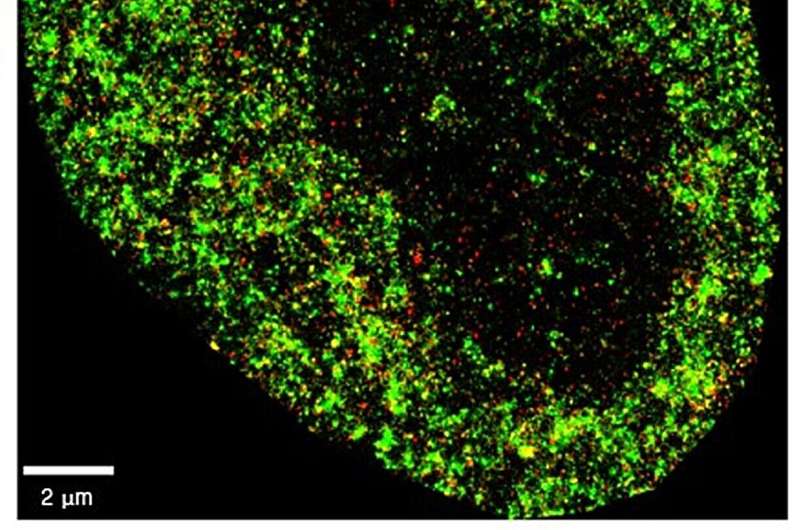
Researchers have developed an artificial intelligence which can differentiate cancer cells from normal cells, as well as detect the very early stages of viral infection inside cells. The findings, published today in a study in the journal Nature Machine Intelligence , pave the way for improved diagnostic techniques and new monitoring strategies for disease. The researchers are from the Centre for Genomic Regulation (CRG), the University of the Basque Country (UPV/EHU), Donostia International Physics Center (DIPC) and the Fundación Biofisica Bizkaia (FBB, located in Biofisika Institute).
The tool, AINU (AI of the NUcleus), scans high-resolution images of cells. The images are obtained with a special microscopy technique called STORM, which creates a picture that captures many finer details than what regular microscopes can see. The high-definition snapshots reveal structures at nanoscale resolution.
A nanometer (nm) is one-billionth of a meter, and a strand of human hair is about 100,000nm wide. The AI can detect rearrangements inside cells as small as 20nm, or 5,000 times smaller than the width of a human hair. These alterations are too small and subtle for human observers to find with traditional methods alone.
"The resolution of these images is powerful enough for our AI to recognize specific patterns and differences with remarkable accuracy, including changes in how DNA is arranged inside cells, helping spot alterations very soon after they occur. We think that, one day, this type of information can buy doctors valuable time to monitor disease, personalize treatments and improve patient outcomes," says ICREA Research Professor Pia Cosma, co-corresponding author of the study and researcher at the Centre for Genomic Regulation in Barcelona.
'Facial recognition' at the molecular level
AINU is a convolutional neural network , a type of AI specifically designed to analyze visual data like images. Examples of convolutional neural networks include AI tools which enable users to unlock smartphones with their face, or others used by self-driving cars to understand and navigate environments by recognizing objects on the road.
In medicine, convolutional neural networks are used to analyze medical images like mammograms or CT scans and identify signs of cancer that might be missed by the human eye. They can also help doctors detect abnormalities in MRI scans or X-ray images, helping make a faster and more accurate diagnosis.
AINU detects and analyzes tiny structures inside cells at the molecular level. The researchers trained the model by feeding it with nanoscale-resolution images of the nucleus of many different types of cells in different states. The model learned to recognize specific patterns in cells by analyzing how nuclear components are distributed and arranged in three-dimensional space.
For example, cancer cells have distinct changes in their nuclear structure compared to normal cells , such as alterations to how their DNA is organized or the distribution of enzymes within the nucleus. After training, AINU could analyze new images of cell nuclei and classify them as cancerous or normal based on these features alone.
The nanoscale resolution of the images enabled the AI to detect changes in a cell's nucleus as soon as one hour after it was infected by the herpes simplex virus type-1. The model could detect the presence of the virus by finding slight differences in how tightly DNA is packed, which happens when a virus starts to alter the structure of the cell's nucleus.
"Our method can detect cells that have been infected by a virus very soon after the infection starts. Normally, it takes time for doctors to spot an infection because they rely on visible symptoms or larger changes in the body. But with AINU, we can see tiny changes in the cell's nucleus right away," says Ignacio Arganda-Carreras, co-corresponding author of the study and Ikerbasque Research Associate at UPV/EHU and affiliated with the FBB-Biofisika Institute and the DIPC in San Sebastián/Donostia.
"Researchers can use this technology to see how viruses affect cells almost immediately after they enter the body, which could help in developing better treatments and vaccines. In hospitals and clinics, AINU could be used to quickly diagnose infections from a simple blood or tissue sample, making the process faster and more accurate," adds Limei Zhong, co-first author of the study and researcher at the Guangdong Provincial People's Hospital (GDPH) in Guangzhou, China.
Laying the groundwork for clinical readiness
The researchers have to overcome important limitations before the technology is ready to be tested or deployed in a clinical setting . For example, STORM images can only be taken with specialized equipment normally only found in biomedical research labs. Setting up and maintaining the imaging systems required by the AI is a significant investment in both equipment and technical expertise.
Another constraint is that STORM imaging typically analyzes only a few cells at a time. For diagnostic purposes, especially in clinical settings where speed and efficiency are crucial, doctors would need to capture many more numbers of cells in a single image to be able to detect or monitor a disease.
"There are many rapid advances in the field of STORM imaging which mean that microscopes may soon be available in smaller or less specialized labs, and eventually, even in the clinic. The limitations of accessibility and throughput are more tractable problems than we previously thought and we hope to carry out preclinical experiments soon," says Dr. Cosma.
Though clinical benefits might be years away, AINU is expected to accelerate scientific research in the short term. The researchers found the technology could identify stem cells with very high precision. Stem cells can develop into any type of cell in the body, an ability known as pluripotency. Pluripotent cells are studied for their potential in helping repair or replace damaged tissues.
AINU can make the process of detecting pluripotent cells quicker and more accurate, helping make stem cell therapies safer and more effective.
"Current methods to detect high-quality stem cells rely on animal testing. However, all our AI model needs to work is a sample that is stained with specific markers that highlight key nuclear features. As well as being easier and faster, it can accelerate stem cell research while contributing to the shift in reducing animal use in science," says Davide Carnevali, first author of the research and researcher at the CRG.
Explore further
Feedback to editors

Multipurpose vaccine shows new promise in the presence of pre-existing immunity
12 hours ago

Researchers develop affordable, rapid blood test for brain cancer

Discovery gives answers to parents of children with rare neurological gene mutation

Human-centered AI tool to improve sepsis management can identify missing information

AI-powered, big data research enhances understanding of systemic vasculitis
13 hours ago

Team develops injectable heart stimulator for emergency situations

Finding epilepsy hotspots before surgery: A faster, non-invasive approach
15 hours ago

Personalized brain stimulation significantly decreases depression symptoms in pilot study

An ancient signaling pathway and 20 years of research offer hope for rare cancer

How mindset could affect the body's response to vaccination
Related stories.

A new way to see viruses in action: Super-resolution microscopy provides a nano-scale look
May 31, 2024

Research harnesses machine learning and imaging to give insight into stem cell behavior
Jul 4, 2024

Observing mammalian cells with superfast soft X-rays
May 24, 2024

Microscopy deep learning predicts viral infections
Jun 21, 2021

AI tool creates 'synthetic' images of cells for enhanced microscopy analysis
Apr 22, 2024

New bioinformatics tool to identify chromosomal alterations in tumor cells
Mar 21, 2024
Recommended for you

New drug combinations could improve therapies for breast cancer, other aggressive cancers

Colorectal cancer: New approach for better efficacy of immunotherapies
17 hours ago
Let us know if there is a problem with our content
Use this form if you have come across a typo, inaccuracy or would like to send an edit request for the content on this page. For general inquiries, please use our contact form . For general feedback, use the public comments section below (please adhere to guidelines ).
Please select the most appropriate category to facilitate processing of your request
Thank you for taking time to provide your feedback to the editors.
Your feedback is important to us. However, we do not guarantee individual replies due to the high volume of messages.
E-mail the story
Your email address is used only to let the recipient know who sent the email. Neither your address nor the recipient's address will be used for any other purpose. The information you enter will appear in your e-mail message and is not retained by Medical Xpress in any form.
Newsletter sign up
Get weekly and/or daily updates delivered to your inbox. You can unsubscribe at any time and we'll never share your details to third parties.
More information Privacy policy
Donate and enjoy an ad-free experience
We keep our content available to everyone. Consider supporting Science X's mission by getting a premium account.
E-mail newsletter
Bubbling, frothing and sloshing: Long-hypothesized plasma instabilities finally observed
Results could aid understanding of how black holes produce vast intergalactic jets.
Whether between galaxies or within doughnut-shaped fusion devices known as tokamaks, the electrically charged fourth state of matter known as plasma regularly encounters powerful magnetic fields, changing shape and sloshing in space. Now, a new measurement technique using protons, subatomic particles that form the nuclei of atoms, has captured details of this sloshing for the first time, potentially providing insight into the formation of enormous plasma jets that stretch between the stars.
Scientists at the U.S. Department of Energy's (DOE) Princeton Plasma Physics Laboratory (PPPL) created detailed pictures of a magnetic field bending outward because of the pressure created by expanding plasma. As the plasma pushed on the magnetic field, bubbling and frothing known as magneto-Rayleigh Taylor instabilities arose at the boundaries, creating structures resembling columns and mushrooms.
Then, as the plasma's energy diminished, the magnetic field lines snapped back into their original positions. As a result, the plasma was compressed into a straight structure resembling the jets of plasma that can stream from ultra-dense dead stars known as black holes and extend for distances many times the size of a galaxy. The results suggest that those jets, whose causes remain a mystery, could be formed by the same compressing magnetic fields observed in this research.
"When we did the experiment and analyzed the data, we discovered we had something big," said Sophia Malko, a PPPL staff research physicist and lead scientist on the paper. "Observing magneto-Rayleigh Taylor instabilities arising from the interaction of plasma and magnetic fields had long been thought to occur but had never been directly observed until now. This observation helps confirm that this instability occurs when expanding plasma meets magnetic fields. We didn't know that our diagnostics would have that kind of precision. Our whole team is thrilled!"
"These experiments show that magnetic fields are very important for the formation of plasma jets," said Will Fox, a PPPL research physicist and principal investigator of the research reported in Physical Review Research. "Now that we might have insight into what generates these jets, we could, in theory, study giant astrophysical jets and learn something about black holes."
PPPL has world-renowned expertise in developing and building diagnostics, sensors that measure properties like density and temperature in plasma in a range of conditions. This achievement is one of several in recent years that illustrates how the Lab is advancing measurement innovation in plasma physics.
Using a new technique to produce unprecedented detail
The team improved a measurement technique known as proton radiography by creating a new variation for this experiment that would allow for extremely precise measurements. To create the plasma, the team shone a powerful laser at a small disk of plastic. To produce protons, they shone 20 lasers at a capsule containing fuel made of varieties of hydrogen and helium atoms. As the fuel heated up, fusion reactions occurred and produced a burst of both protons and intense light known as X-rays.
The team also installed a sheet of mesh with tiny holes near the capsule. As the protons flowed through the mesh, the outpouring was separated into small, separate beams that were bent because of the surrounding magnetic fields. By comparing the distorted mesh image to an undistorted image produced by X-rays, the team could understand how the magnetic fields were pushed around by the expanding plasma, leading to whirl-like instabilities at the edges.
"Our experiment was unique because we could directly see the magnetic field changing over time," Fox said. "We could directly observe how the field gets pushed out and responds to the plasma in a type of tug of war."
Diversifying a research portfolio
The findings exemplify how PPPL is expanding its focus to include research focused on high energy density (HED) plasma. Such plasmas, like the one created in this experiment's fuel capsule, are hotter and denser than those used in fusion experiments. "HED plasma is an exciting area of growth for plasma physics," Fox said. "This work is part of PPPL's efforts to advance this field. The results show how the Laboratory can create advanced diagnostics to give us new insights into this type of plasma, which can be used in laser fusion devices, as well as in techniques that use HED plasma to create radiation for microelectronics manufacturing."
"PPPL has an enormous amount of knowledge and experience in magnetized plasmas that can contribute to the field of laser-produced HED plasmas and help make significant contributions," Fox said.
"HED science is complex, fascinating and key to understanding a wide range of phenomena," said Laura Berzak Hopkins, PPPL's associate laboratory director for strategy and partnerships and deputy chief research officer. "It's incredibly challenging to both generate these conditions in a controlled manner and develop advanced diagnostics for precision measurements. These exciting results demonstrate the impact of integrating PPPL's breadth of technical expertise with innovative approaches."
More experiments and better simulations
The researchers plan to work on future experiments that will help improve models of expanding plasma. "Scientists have assumed that in these situations, density and magnetism vary directly, but it turns out that that's not true," Malko said.
"Now that we have measured these instabilities very accurately, we have the information we need to improve our models and potentially simulate and understand astrophysical jets to a higher degree than before," Malko said. "It's interesting that humans can make something in a laboratory that usually exists in space."
Collaborators included researchers from the University of California-Los Angeles, the Sorbonne University, Princeton University and the University of Michigan. The research was funded by the DOE's Laboratory-Directed Research and Development program under contract number DE-AC02-09CH11466. The experiment was conducted using the University of Rochester's Omega Laser Facility under DOE/National Nuclear Security Administration contract number DE-NA0003856.
- Astrophysics
- Solar Flare
- Black Holes
- Nuclear Energy
- Medical Technology
- Interstellar medium
- Van Allen radiation belt
- Solar flare
- Nucleosynthesis
- Red supergiant star
- Magnetic resonance imaging
Story Source:
Materials provided by DOE/Princeton Plasma Physics Laboratory . Original written by Raphael Rosen. Note: Content may be edited for style and length.
Journal Reference :
- S. Malko, D. B. Schaeffer, W. Yao, V. Valenzuela-Villaseca, C. Johnson, G. Fiksel, A. Ciardi, W. Fox. Observation of a magneto-Rayleigh-Taylor instability in magnetically collimated plasma jets . Physical Review Research , 2024; 6 (2) DOI: 10.1103/PhysRevResearch.6.023330
Cite This Page :
Explore More
- Six New Rogue Worlds: Star Birth Clues
- Ancient Climate Change and Fossilized Shells
- How Black Holes Produce Vast Intergalactic Jets
- Hidden Magmatism On the Moon
- Black Holes in High Res
- Your Favorite Film Genres and Your Brain
- Annual Breast Cancer Screening for Women Over 40
- Pregnancy After 'Permanent Sterilization'
- Climate: Coastal Cities Must Adapt Faster
- No Crisis in Cosmology, After All
Trending Topics
Strange & offbeat.

IMAGES
VIDEO
COMMENTS
At UWA, Medical Physics research is strongly aligned with improving treatment and diagnostic precision using localized and minimally invasive techniques that are aimed to improve patient outcomes.
Medical Physics is a journal of global scope and reach. By publishing in Medical Physics your research will reach an international, multidisciplinary audience including practicing medical physicists as well as physics- and engineering based translational scientists. We work closely with authors of promising articles to improve their quality.
Medical Physics Research The mission of physics research is to develop new techniques and investigate new methodologies to continuously improve treatment planning and evaluation methods, target localization and verification accuracy and treatment delivery precision.
BMC Medical Physics is an open access journal publishing original peer-reviewed research articles in all aspects of medical physics, including radiobiology, ...
Explore the latest full-text research PDFs, articles, conference papers, preprints and more on MEDICAL PHYSICS. Find methods information, sources, references or conduct a literature review on ...
Medical physics is a constantly evolving field of physics applied to healthcare, with the objective of ensuring safety, quality and technology implementation in diagnostic and therapeutic medicine.
This month we examine examples of how advances from different areas of physics can lead to medical applications.
Medical physics is a very well-established field where advances tend to be of technical nature. However, physicists from other areas of physics can make unexpected contributions. New ideas ...
It has been our privilege to witness and facilitate dissemination of cutting-edge medical physics research, which encompasses a broad and ever-enlarging range of topics, clinical applications, and methodologies.
Health Physics, first published in 1958, provides the latest research to a wide variety of radiation safety professionals including health physicists, nuclear chemists, medical physicists, and radiation safety officers with interests in nuclear and radiation science.
Our faculty are drawn from 5 departments: Radiology, Radiation Oncology, Radiation Safety, Biomedical Engineering, and Physics. In addition, each faculty affiliates with one or more of the program's four tracks. Research Highlights from Duke Medical Physics Program on Vimeo. The tracks are shown below with representative topics of research ...
Ultimately, it is about "partnership" of medical physics and medicine, and imaging is a playground with little confrontation. Our journal, Frontiers in Physics—Medical Physics and Imaging, seeks to provide a forum that captures the bespoke cross-disciplinary engagement of medical physicists and imaging experts.
AAPM Reports are technical reports on specific topics that are prepared by experts drawn from the AAPM membership who work together in Task Groups under the guidance of the AAPM Councils and Committees. All AAPM reports are published on-line with open-access without subscription.
The Oficial Voice of the AAPM as well as abstracts from the AAPM Annual Meeting. Additionally, regular features like Point/Counterpoint discussions of controversial issues and Future of Medical Physics papers ar itals, clinics, universities, and research centers. They a Diagnostic Radiology Therapeutic Radiology Nuclear Medicine
Explores research on theoretical, experimental, and computational techniques relating to the expanding, interdisciplinary field of bio-medical physics.
The panel discussion will explore opportunities and vistas in medical physics research and practice, medical imaging, teaching medical physics to undergraduates, and medical physics curricula as a recruiting tool for physics departments.
The principles and methods of applied physics have long been applied to Medicine for the design of diagnostic and therapeutic techniques through the use of ionizing and non-ionizing radiation. Medical Physics covers all areas of applied physics research dealing with the prevention, diagnosis, and treatment of human diseases.
Message from Certificate Program Director The Medical Physics Certificate Program (MPCP) is a rigorous two-year (CAMPEP-accreditation pending) didactic training program, meticulously designed and administered by the Departments of Radiation Oncology & Radiology at Stanford University School of Medicine. The curriculum covers essential medical physics topics, aligning with AAPM guidelines and ...
The American Association of Physicists in Medicine is a member society concerned with the topics of medical physics, radiation oncology, imaging physics, health physics, hospital physics, medical radiation, physics careers, ionizing radiation, brachytherapy and diagnostic imaging.
The field of medical physics has struggled with the role of research in recent years, as professional interests have dominated its growth toward clinical service. This article focuses on the subset of medical physics programs within academic medical centers and how a refocused academic mission withi …
Current research. Both the MSc and PhD programs in Medical Physics give students the opportunity to engage in impactful and innovative research, supervised by leading faculty in medical imaging and radiation oncology physics. The majority of thesis supervisors are certified clinical medical physicists, which means that research projects are ...
Some common research topics in Medical Physics include radiation therapy, medical imaging, nuclear medicine, biological effects of radiation, medical device development, and radiation safety. 3.
The "Split Curriculum". While many American medical schools are decreasing the extent to which medical students study basic science, advances in molecular medicine, and research in general, Stanford created a curriculum - the "Split Curriculum"- that restores vigor to the basic courses and provides opportunities to engage in other scholarly activities available at Stanford University.
Researchers have developed an artificial intelligence which can differentiate cancer cells from normal cells, as well as detect the very early stages of viral infection inside cells. The findings ...
The research team has developed a magnetic noise suppression method based on magnetic noise self-compensation effects, and experimentally verified it in a mixed system of potassium (K) and 3 He gases.
Scientists have observed new details of how plasma interacts with magnetic fields, potentially providing insight into the formation of enormous plasma jets that stretch between the stars.